9J1W | pdb_00009j1w

Endogenous dihydrolipoamide acetyltransferase (E2) core of pyruvate dehydrogenase complex from pig heart

  • Classification: TRANSFERASE
  • Organism(s): Sus scrofa
  • Mutation(s): No 

  • Deposited: 2024-08-05 Released: 2025-02-05 
  • Deposition Author(s): Wang, C., Zhang, X., Chang, Y.J.
  • Funding Organization(s): National Natural Science Foundation of China (NSFC)

Experimental Data Snapshot

  • Method: ELECTRON MICROSCOPY
  • Resolution: 3.20 Å
  • Aggregation State: PARTICLE 
  • Reconstruction Method: SINGLE PARTICLE 

wwPDB Validation 3D Report Full Report

Validation slider image for 9J1W

This is version 1.0 of the entry. See complete history

Literature

Dynamics of the mammalian pyruvate dehydrogenase complex revealed by in-situ structural analysis.

Wang, C.Ma, C.Xu, Y.Chang, S.Wu, H.Yan, C.Chen, J.Wu, Y.An, S.Xu, J.Han, Q.Jiang, Y.Jiang, Z.Chu, X.Gao, H.Zhang, X.Chang, Y.

(2025) Nat Commun 16: 917-917

  • DOI: https://doi.org/10.1038/s41467-025-56171-8
  • Primary Citation Related Structures: 
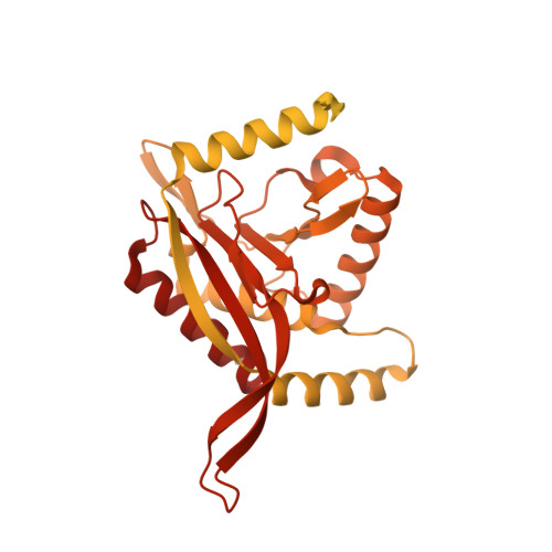
    9J1W

  • PubMed Abstract: 

    The multi-enzyme pyruvate dehydrogenase complex (PDHc) links glycolysis to the citric acid cycle and plays vital roles in metabolism, energy production, and cellular signaling. Although all components have been individually characterized, the intact PDHc structure remains unclear, hampering our understanding of its composition and dynamical catalytic mechanisms. Here, we report the in-situ architecture of intact mammalian PDHc by cryo-electron tomography. The organization of peripheral E1 and E3 components varies substantially among the observed PDHcs, with an average of 21 E1 surrounding each PDHc core, and up to 12 E3 locating primarily along the pentagonal openings. In addition, we observed dynamic interactions of the substrate translocating lipoyl domains (LDs) with both E1 and E2, and the interaction interfaces were further analyzed by molecular dynamics simulations. By revealing intrinsic dynamics of PDHc peripheral compositions, our findings indicate a distinctive activity regulation mechanism, through which the number of E1, E3 and functional LDs may be coordinated to meet constantly changing demands of metabolism.


  • Organizational Affiliation
    • Department of Pathology of Sir Run Run Shaw Hospital and Department of Biophysics, Zhejiang University School of Medicine, Hangzhou, Zhejiang, China.

Macromolecule Content 

  • Total Structure Weight: 4,155.51 kDa 
  • Atom Count: 105,420 
  • Modeled Residue Count: 13,680 
  • Deposited Residue Count: 38,820 
  • Unique protein chains: 1

Macromolecules

Find similar proteins by:|  3D Structure
Entity ID: 1
MoleculeChains  Sequence LengthOrganismDetailsImage
Acetyltransferase component of pyruvate dehydrogenase complex647Sus scrofaMutation(s): 0 
EC: 2.3.1.12
UniProt
Find proteins for A0A5G2QLQ1 (Sus scrofa)
Explore A0A5G2QLQ1 
Go to UniProtKB:  A0A5G2QLQ1
Entity Groups
Sequence Clusters30% Identity50% Identity70% Identity90% Identity95% Identity100% Identity
UniProt GroupA0A5G2QLQ1
Sequence Annotations
Expand
Reference Sequence

Experimental Data & Validation

Experimental Data

  • Method: ELECTRON MICROSCOPY
  • Resolution: 3.20 Å
  • Aggregation State: PARTICLE 
  • Reconstruction Method: SINGLE PARTICLE 

Structure Validation

View Full Validation Report


Entry History 

& Funding Information

Deposition Data


Funding OrganizationLocationGrant Number
National Natural Science Foundation of China (NSFC)China2018YFA0507700
National Natural Science Foundation of China (NSFC)China32200980

Revision History  (Full details and data files)

  • Version 1.0: 2025-02-05
    Type: Initial release