


Crystal structure of CTB10-PE3.0-(R)-1g
Fu, K., Rao, Y.J.To be published.
Experimental Data Snapshot
Starting Model: experimental
View more details
Entity ID: 1 | |||||
|---|---|---|---|---|---|
| Molecule | Chains | Sequence Length | Organism | Details | Image |
| CTB10 | 141 | Cercospora sp. JNU001 | Mutation(s): 2 | 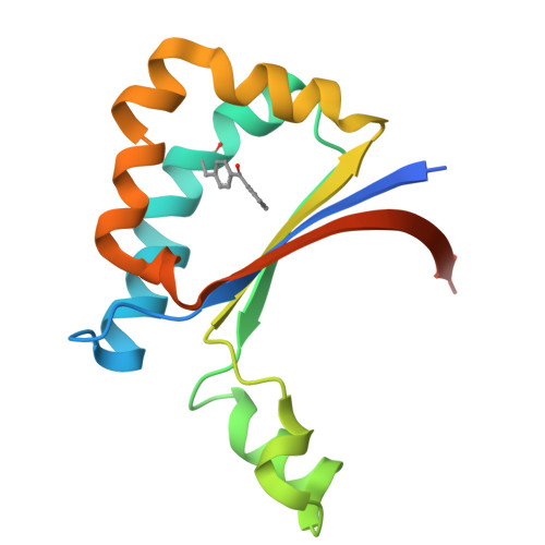 | |
UniProt | |||||
Entity Groups | |||||
| Sequence Clusters | 30% Identity50% Identity70% Identity90% Identity95% Identity100% Identity | ||||
| UniProt Group | A0A977K7H6 | ||||
Sequence AnnotationsExpand | |||||
Reference Sequence | |||||
| Ligands 3 Unique | |||||
|---|---|---|---|---|---|
| ID | Chains | Name / Formula / InChI Key | 2D Diagram | 3D Interactions | |
| A1EAB (Subject of Investigation/LOI) Download:Ideal Coordinates CCD File | I [auth A] K [auth C] L [auth E] N [auth F] O [auth G] | 2-[(3-cyanophenyl)methyl]-5,5-dimethyl-hexa-2,3-dienamide C16 H18 N2 O OISKZRHWMMCOBF-ZETCQYMHSA-N |  | ||
| PEG Download:Ideal Coordinates CCD File | J [auth B] | DI(HYDROXYETHYL)ETHER C4 H10 O3 MTHSVFCYNBDYFN-UHFFFAOYSA-N |  | ||
| GOL Download:Ideal Coordinates CCD File | M [auth F], P [auth H], Q [auth H] | GLYCEROL C3 H8 O3 PEDCQBHIVMGVHV-UHFFFAOYSA-N |  | ||
| Modified Residues 1 Unique | |||||
|---|---|---|---|---|---|
| ID | Chains | Type | Formula | 2D Diagram | Parent |
| PBF Query on PBF | A, B, C, D, E A, B, C, D, E, F, G, H | L-PEPTIDE LINKING | C16 H15 N O3 |  | PHE |
| Length ( Å ) | Angle ( ˚ ) |
|---|---|
| a = 39.13 | α = 90 |
| b = 163.71 | β = 93.01 |
| c = 87.408 | γ = 90 |
| Software Name | Purpose |
|---|---|
| PHENIX | refinement |
| XDS | data reduction |
| Aimless | data scaling |
| MOLREP | phasing |
| Funding Organization | Location | Grant Number |
|---|---|---|
| Other private | China | 2021YFC2102700 |
| Other private | China | BK20202002 |
| Other private | China | KYCX20_1812 |
| Other private | China | KYCX20_1816 |


