


Crystal structure of human NAMPT complexed with AMP
Wang, G., Wu, C.To be published.
Experimental Data Snapshot
Starting Model: experimental
View more details
Entity ID: 1 | |||||
|---|---|---|---|---|---|
| Molecule | Chains | Sequence Length | Organism | Details | Image |
| Nicotinamide phosphoribosyltransferase | 497 | Homo sapiens | Mutation(s): 0 Gene Names: NAMPT, PBEF, PBEF1 EC: 2.4.2.12 | 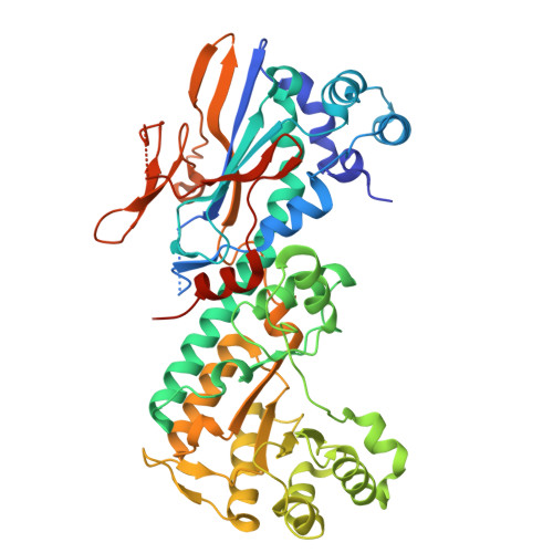 | |
UniProt & NIH Common Fund Data Resources | |||||
PHAROS: P43490 GTEx: ENSG00000105835 | |||||
Entity Groups | |||||
| Sequence Clusters | 30% Identity50% Identity70% Identity90% Identity95% Identity100% Identity | ||||
| UniProt Group | P43490 | ||||
Sequence AnnotationsExpand | |||||
Reference Sequence | |||||
| Ligands 3 Unique | |||||
|---|---|---|---|---|---|
| ID | Chains | Name / Formula / InChI Key | 2D Diagram | 3D Interactions | |
| AMP (Subject of Investigation/LOI) Download:Ideal Coordinates CCD File | E [auth A], I [auth B], L [auth C], P [auth D] | ADENOSINE MONOPHOSPHATE C10 H14 N5 O7 P UDMBCSSLTHHNCD-KQYNXXCUSA-N |  | ||
| PO4 Download:Ideal Coordinates CCD File | F [auth A] G [auth A] J [auth B] K [auth B] M [auth C] | PHOSPHATE ION O4 P NBIIXXVUZAFLBC-UHFFFAOYSA-K |  | ||
| MG Download:Ideal Coordinates CCD File | H [auth A], O [auth C], S [auth D] | MAGNESIUM ION Mg JLVVSXFLKOJNIY-UHFFFAOYSA-N |  | ||
| Length ( Å ) | Angle ( ˚ ) |
|---|---|
| a = 86.17 | α = 90 |
| b = 93.374 | β = 90 |
| c = 241.942 | γ = 90 |
| Software Name | Purpose |
|---|---|
| PHENIX | refinement |
| HKL-2000 | data reduction |
| HKL-2000 | data scaling |
| PHENIX | phasing |
| Funding Organization | Location | Grant Number |
|---|---|---|
| National Natural Science Foundation of China (NSFC) | China | 81872874 |
| National Natural Science Foundation of China (NSFC) | China | 91949101 |


