9IJD | pdb_00009ijd

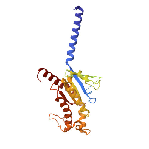
Carazolol-activated human beta3 adrenergic receptor


Experimental Data Snapshot

  • Method: ELECTRON MICROSCOPY
  • Resolution: 2.76 Å
  • Aggregation State: PARTICLE 
  • Reconstruction Method: SINGLE PARTICLE 

wwPDB Validation 3D Report Full Report

Validation slider image for 9IJD

This is version 1.3 of the entry. See complete history

Literature

Molecular Mechanism of the beta 3 AR Agonist Activity of a beta-Blocker.

Zheng, S.Zhang, S.Dai, S.Chen, K.Gao, K.Sun, X.Lin, B.Liu, X.

(2024) Chempluschem 89: e202400288-e202400288

  • DOI: https://doi.org/10.1002/cplu.202400288
  • Primary Citation Related Structures: 
    9IJD, 9IJE

  • PubMed Abstract: 

    Development of subtype-selective drugs for G protein-coupled receptors poses a significant challenge due to high similarity between subtypes, as exemplified by the three β-adrenergic receptors (βARs). The β 3 AR agonists show promise for treating the overactive bladder or preterm birth, but their potential is hindered by off-target activation of β 1 AR and β 2 AR. Interestingly, several β-blockers, which are antagonists of the β 1 ARs and β 2 ARs, have been reported to exhibit agonist activity at the β 3 AR. However, the molecular mechanism remains elusive. Understanding the underlying mechanism should facilitate the development of β 3 AR agonists with improved selectivity and reduced off-target effects. In this work, we determined the structures of human β 3 AR in complex with the endogenous agonist epinephrine or with a synthetic β 3 AR agonist carazolol, which is also a high-affinity β-blocker. Structure comparison, mutagenesis studies and molecular dynamics simulations revealed that the differences on the flexibility of D 3.32 directly contribute to carazolol's distinct activities as an antagonist for the β 2 AR and an agonist for the β 3 AR. The process is also indirectly influenced by the extracellular loops (ECL), especially ECL1. Taken together, these results provide key guidance for development of selective β 3 AR agonists, paving the way for new therapeutic opportunities.


  • Organizational Affiliation
    • State Key Laboratory of Membrane Biology, Tsinghua-Peking Center for Life Sciences, School of Pharmaceutical Sciences, Tsinghua University, Beijing, 100084, P. R. China.

Macromolecule Content 

  • Total Structure Weight: 161.5 kDa 
  • Atom Count: 9,371 
  • Modeled Residue Count: 1,227 
  • Deposited Residue Count: 1,460 
  • Unique protein chains: 6

Macromolecules

Find similar proteins by:|  3D Structure
Entity ID: 1
MoleculeChains  Sequence LengthOrganismDetailsImage
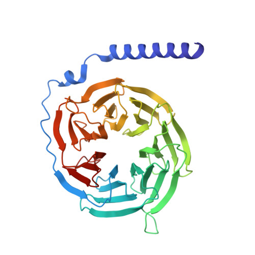
Guanine nucleotide-binding protein G(I)/G(S)/G(T) subunit beta-1A [auth B]340Rattus norvegicusMutation(s): 0 
Gene Names: Gnb1
UniProt
Find proteins for P54311 (Rattus norvegicus)
Explore P54311 
Go to UniProtKB:  P54311
Entity Groups
Sequence Clusters30% Identity50% Identity70% Identity90% Identity95% Identity100% Identity
UniProt GroupP54311
Sequence Annotations
Expand
Reference Sequence
Find similar proteins by:|  3D Structure
Entity ID: 2
MoleculeChains  Sequence LengthOrganismDetailsImage
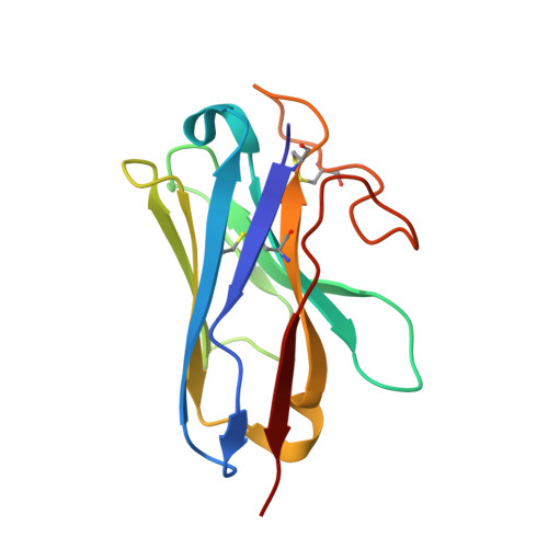
Camelid antibody VHH fragmentB [auth N]128Lama glamaMutation(s): 0 
Entity Groups
Sequence Clusters30% Identity50% Identity70% Identity90% Identity95% Identity100% Identity
Sequence Annotations
Expand
Reference Sequence
Find similar proteins by:|  3D Structure
Entity ID: 3
MoleculeChains  Sequence LengthOrganismDetailsImage
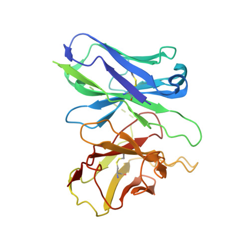
Single-chain Fv16C [auth D]246Homo sapiensMutation(s): 0 
Entity Groups
Sequence Clusters30% Identity50% Identity70% Identity90% Identity95% Identity100% Identity
Sequence Annotations
Expand
Reference Sequence
Find similar proteins by:|  3D Structure
Entity ID: 4
MoleculeChains  Sequence LengthOrganismDetailsImage
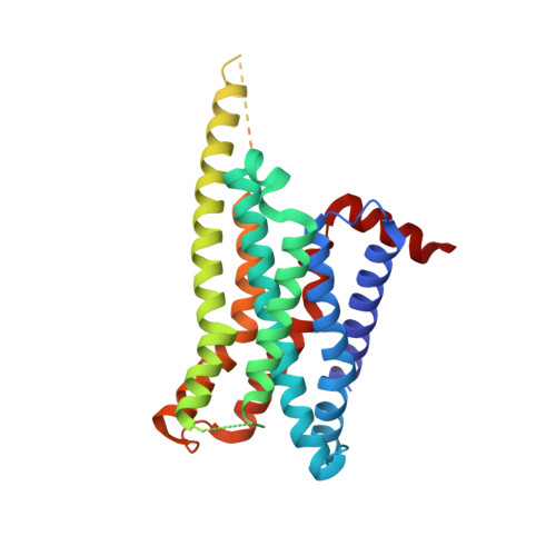
Beta-3 adrenergic receptorD [auth R]321Homo sapiensMutation(s): 0 
Gene Names: ADRB3ADRB3RB3AR
Membrane Entity: Yes 
UniProt & NIH Common Fund Data Resources
Find proteins for P13945 (Homo sapiens)
Explore P13945 
Go to UniProtKB:  P13945
GTEx:  ENSG00000188778 
Entity Groups
Sequence Clusters30% Identity50% Identity70% Identity90% Identity95% Identity100% Identity
UniProt GroupP13945
Sequence Annotations
Expand
Reference Sequence
Find similar proteins by:|  3D Structure
Entity ID: 5
MoleculeChains  Sequence LengthOrganismDetailsImage
Guanine nucleotide-binding protein G(s) subunit alpha isoforms shortE [auth A]373Bos taurusMutation(s): 0 
Gene Names: GNASGNAS1
EC: 3.6.5
Membrane Entity: Yes 
UniProt
Find proteins for P04896 (Bos taurus)
Explore P04896 
Go to UniProtKB:  P04896
Entity Groups
Sequence Clusters30% Identity50% Identity70% Identity90% Identity95% Identity100% Identity
UniProt GroupP04896
Sequence Annotations
Expand
Reference Sequence
Find similar proteins by:|  3D Structure
Entity ID: 6
MoleculeChains  Sequence LengthOrganismDetailsImage
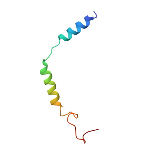
Guanine nucleotide-binding protein G(I)/G(S)/G(O) subunit gamma-2F [auth C]52Bos taurusMutation(s): 0 
Gene Names: GNG2
Membrane Entity: Yes 
UniProt
Find proteins for P63212 (Bos taurus)
Explore P63212 
Go to UniProtKB:  P63212
Entity Groups
Sequence Clusters30% Identity50% Identity70% Identity90% Identity95% Identity100% Identity
UniProt GroupP63212
Sequence Annotations
Expand
Reference Sequence

Small Molecules

Ligands 1 Unique
IDChains Name / Formula / InChI Key2D Diagram3D Interactions
CAU
(Subject of Investigation/LOI)

Query on CAU


Download:Ideal Coordinates CCD File
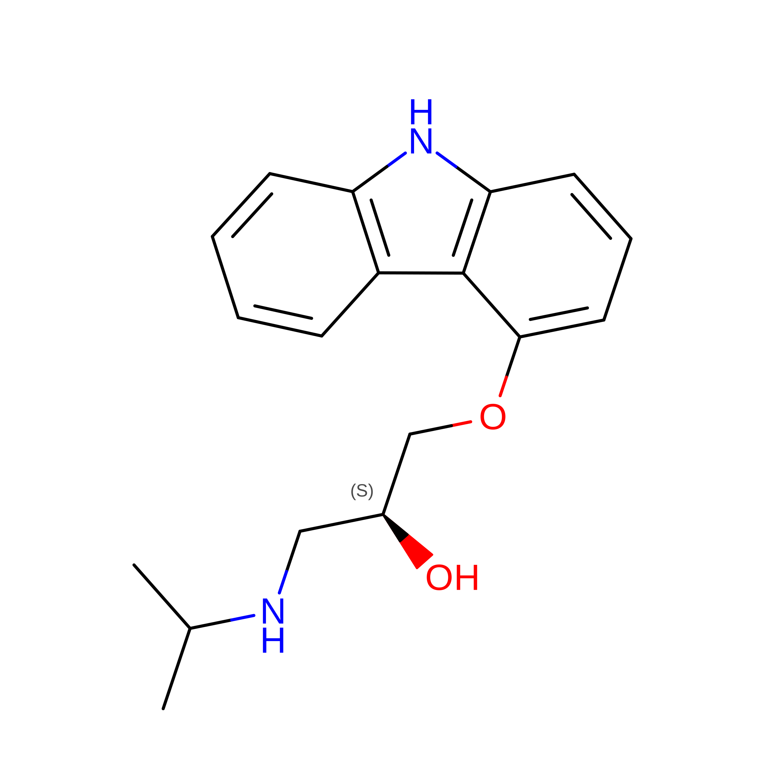
G [auth R](2S)-1-(9H-Carbazol-4-yloxy)-3-(isopropylamino)propan-2-ol
C18 H22 N2 O2
BQXQGZPYHWWCEB-ZDUSSCGKSA-N

Experimental Data & Validation

Experimental Data

  • Method: ELECTRON MICROSCOPY
  • Resolution: 2.76 Å
  • Aggregation State: PARTICLE 
  • Reconstruction Method: SINGLE PARTICLE 
EM Software:
TaskSoftware PackageVersion
MODEL REFINEMENTPHENIX

Structure Validation

View Full Validation Report


Entry History 

& Funding Information

Deposition Data


Funding OrganizationLocationGrant Number
National Natural Science Foundation of China (NSFC)China32122041

Revision History  (Full details and data files)

  • Version 1.0: 2024-08-21
    Type: Initial release
  • Version 1.1: 2024-11-06
    Changes: Data collection, Structure summary
  • Version 1.2: 2024-12-25
    Changes: Data collection, Database references
  • Version 1.3: 2025-07-23
    Changes: Data collection