9I7O | pdb_00009i7o

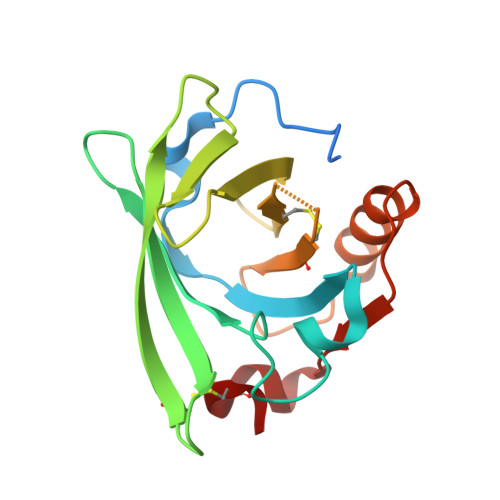
Beta-lactoglobulin complex with retinol


Experimental Data Snapshot

  • Method: X-RAY DIFFRACTION
  • Resolution: 2.00 Å
  • R-Value Free: 
    0.197 (Depositor), 0.197 (DCC) 
  • R-Value Work: 
    0.176 (Depositor), 0.176 (DCC) 
  • R-Value Observed: 
    0.177 (Depositor) 

Starting Model: experimental
View more details

wwPDB Validation 3D Report Full Report

Validation slider image for 9I7O

Ligand Structure Quality Assessment 


This is version 1.1 of the entry. See complete history. 

Literature

New solid formulations of retinol protected at the molecular level by the whey protein beta-lactoglobulin.

Figueiredo, M., Sarkany, Z., Pereira, P.J.B., Rocha, F., Martins, P.M.

(2026) Food Chem 526: 150783-150783

  • DOI: https://doi.org/10.1016/j.foodchem.2026.150783
  • Primary Citation Related Structures: 
    9I7N, 9I7O

  • PubMed Abstract: 

    The fat-soluble vitamin retinol plays a critical role in vision, immune function, and skin health, with deficiencies linked to severe health issues, including blindness and increased risk of infection. Despite its importance to the food industry, it is poorly soluble in water and degrades rapidly when exposed to light, heat, and oxygen, limiting its use as a nutraceutical. This study introduces a novel solid formulation of retinol encapsulated by the whey protein β-lactoglobulin (β-LG) to enhance stability for food fortification applications. Retinol forms molecular complexes with β-LG, which are dried into a solid powder, minimizing degradation. Encapsulation within the β-LG calyx protects retinol at the molecular level, as confirmed by fluorescence quenching, mass spectrometry, and X-ray crystallography. This formulation is able to retain retinol integrity throughout more than 5 months of extended storage, which benefits the practical application of the vitamin, addressing health needs while ensuring stability during processing and storage.


  • Organizational Affiliation: 
    • LEPABE, ALiCE, Faculty of Engineering, University of Porto, Rua Dr. Roberto Frias, 4200-465 Porto, Portugal; i3S - Instituto de Investigação e Inovação em Saúde, Universidade do Porto, Rua Alfredo Allen 208, 4200-135 Porto, Portugal.

Macromolecule Content 

  • Total Structure Weight: 20.28 kDa 
  • Atom Count: 1,303 
  • Modeled Residue Count: 159 
  • Deposited Residue Count: 180 
  • Unique protein chains: 1

Macromolecules

Find similar proteins by:|  3D Structure
Entity ID: 1
MoleculeChains  Sequence LengthOrganismDetailsImage
Beta-lactoglobulin178Bos taurusMutation(s): 2 
UniProt
Find proteins for P02754 (Bos taurus)
Explore P02754 
Go to UniProtKB:  P02754
Entity Groups
Sequence Clusters30% Identity50% Identity70% Identity90% Identity95% Identity100% Identity
UniProt GroupP02754
Sequence Annotations
Expand
Reference Sequence

Small Molecules

Ligands 1 Unique
IDChains Name / Formula / InChI Key2D Diagram3D Interactions
RTL
(Subject of Investigation/LOI)

Query on RTL


Download:Ideal Coordinates CCD File
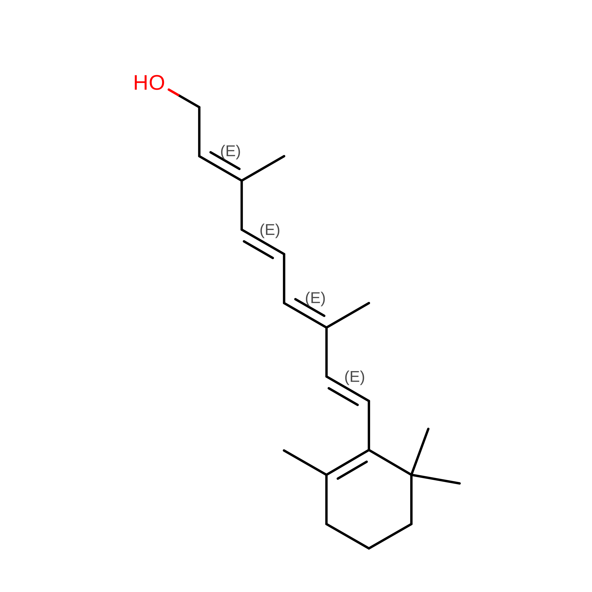
B [auth A]RETINOL
C20 H30 O
FPIPGXGPPPQFEQ-OVSJKPMPSA-N
Binding Affinity Annotations 
IDSourceBinding Affinity
RTL BindingDB:  9I7O Kd: 36 (nM) from 1 assay(s)

Experimental Data & Validation

Experimental Data

  • Method: X-RAY DIFFRACTION
  • Resolution: 2.00 Å
  • R-Value Free:  0.197 (Depositor), 0.197 (DCC) 
  • R-Value Work:  0.176 (Depositor), 0.176 (DCC) 
  • R-Value Observed: 0.177 (Depositor) 
Space Group: P 32 2 1
Unit Cell:
Length ( Å )Angle ( ˚ )
a = 53.907α = 90
b = 53.907β = 90
c = 112.579γ = 120
Software Package:
Software NamePurpose
PHENIXrefinement
XDSdata reduction
Aimlessdata scaling
PHASERphasing

Structure Validation

View Full Validation Report


Ligand Structure Quality Assessment 


Entry History 

& Funding Information

Deposition Data


Funding OrganizationLocationGrant Number
Fundacao para a Ciencia e a TecnologiaPortugal2023.00318.BD

Revision History  (Full details and data files)

  • Version 1.0: 2026-02-18
    Type: Initial release
  • Version 1.1: 2026-09-09
    Changes: Database references