9I63 | pdb_00009i63

Synthetic Human Saposin D glycoprotein


Experimental Data Snapshot

  • Method: X-RAY DIFFRACTION
  • Resolution: 1.65 Å
  • R-Value Free: 
    0.201 (Depositor), 0.211 (DCC) 
  • R-Value Work: 
    0.161 (Depositor), 0.176 (DCC) 

Starting Model: experimental
View more details

wwPDB Validation 3D Report Full Report

Validation slider image for 9I63

This is version 1.0 of the entry. See complete history

Literature

Synthetic Glycoforms Reveal Carbohydrate-Dependent Bioactivity of Human Saposin D.

Graf, C.G.F.Schulz, C.Schmalzlein, M.Heinlein, C.Monnich, M.Perkams, L.Puttner, M.Boos, I.Hessefort, M.Lombana Sanchez, J.N.Weyand, M.Steegborn, C.Breiden, B.Ross, K.Schwarzmann, G.Sandhoff, K.Unverzagt, C.

(2017) Angew Chem Int Ed Engl 56: 5252-5257

  • DOI: https://doi.org/10.1002/anie.201701362
  • Primary Citation Related Structures: 
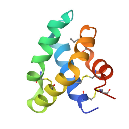
    9I63

  • PubMed Abstract: 

    The main glycoforms of the hydrophobic lysosomal glycoprotein saposin D (SapD) were synthesized by native chemical ligation. An approach for the challenging solid-phase synthesis of the fragments was developed. Three SapD glycoforms were obtained following a general and robust refolding and purification protocol. A crystal structure of one glycoform confirmed its native structure and disulfide pattern. Functional assays revealed that the lipid-binding properties of three SapD glycoforms are highly affected by the single sugar moiety of SapD showing a dependency of the size and the type of N-glycan.


  • Organizational Affiliation
    • Bioorg. Chemie, Gebäude NWI, Universität Bayreuth, 95440, Bayreuth, Germany.

Macromolecule Content 

  • Total Structure Weight: 18.33 kDa 
  • Atom Count: 1,501 
  • Modeled Residue Count: 156 
  • Deposited Residue Count: 164 
  • Unique protein chains: 1

Macromolecules

Find similar proteins by:|  3D Structure
Entity ID: 1
MoleculeChains  Sequence LengthOrganismDetailsImage
Prosaposin
A, B
82Homo sapiensMutation(s): 1 
UniProt & NIH Common Fund Data Resources
Find proteins for P07602 (Homo sapiens)
Explore P07602 
Go to UniProtKB:  P07602
PHAROS:  P07602
GTEx:  ENSG00000197746 
Entity Groups
Sequence Clusters30% Identity50% Identity70% Identity90% Identity95% Identity100% Identity
UniProt GroupP07602
Sequence Annotations
Expand
Reference Sequence

Small Molecules

Ligands 1 Unique
IDChains Name / Formula / InChI Key2D Diagram3D Interactions
SO4

Query on SO4


Download:Ideal Coordinates CCD File
C [auth B]SULFATE ION
O4 S
QAOWNCQODCNURD-UHFFFAOYSA-L
Modified Residues  1 Unique
IDChains TypeFormula2D DiagramParent
NLE
Query on NLE
A, B
L-PEPTIDE LINKINGC6 H13 N O2LEU

Experimental Data & Validation

Experimental Data

  • Method: X-RAY DIFFRACTION
  • Resolution: 1.65 Å
  • R-Value Free:  0.201 (Depositor), 0.211 (DCC) 
  • R-Value Work:  0.161 (Depositor), 0.176 (DCC) 
Space Group: P 1 21 1
Unit Cell:
Length ( Å )Angle ( ˚ )
a = 40.44α = 90
b = 65.68β = 114.498
c = 42.47γ = 90
Software Package:
Software NamePurpose
REFMACrefinement
XDSdata reduction
XSCALEdata scaling
PHASERphasing

Structure Validation

View Full Validation Report


Entry History 

& Funding Information

Deposition Data


Funding OrganizationLocationGrant Number
German Research Foundation (DFG)GermanyUN63/5-1

Revision History  (Full details and data files)

  • Version 1.0: 2025-04-09
    Type: Initial release