9HYW | pdb_00009hyw

Cryo-EM structure of ATTRv-F64S amyloid fibril from patient skin tissue.


Experimental Data Snapshot

  • Method: ELECTRON MICROSCOPY
  • Resolution: 2.80 Å
  • Aggregation State: HELICAL ARRAY 
  • Reconstruction Method: SINGLE PARTICLE 

Starting Model: experimental
View more details

wwPDB Validation 3D Report Full Report

Validation slider image for 9HYW

This is version 1.1 of the entry. See complete history

Literature

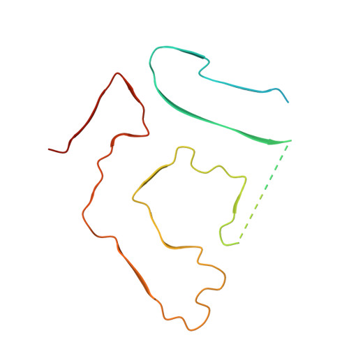
Structure of ATTRv-F64S fibrils isolated from skin tissue of a living patient.

Yu, J.Zhang, X.Pinton, S.Vacchi, E.Cavalli, A.Pecoraro, M.Melli, G.Boland, A.

(2025) Nat Commun 17: 781-781

  • DOI: https://doi.org/10.1038/s41467-025-67457-2
  • Primary Citation Related Structures: 
    9HYW

  • PubMed Abstract: 

    Amyloid transthyretin-derived (ATTR) amyloidosis is a degenerative, systemic disease characterized by transthyretin fibril deposition in organs like the heart, kidneys, liver, and skin. In this study, we report the cryo-EM structure of transthyretin fibrils isolated from skin tissue of a living patient carrying a rare genetic mutation (ATTRv F64S). The structure adopts a highly conserved fold previously observed in other ATTR fibrils from various tissues or different genetic variants. Mass spectrometry was used to evaluate fibril content and to identify common post-translational modifications. The structural consistency between ATTR filaments from different tissues or patients validates non-invasive skin biopsy as a diagnostic tool.


  • Organizational Affiliation
    • Department of Molecular and Cellular Biology, University of Geneva, Geneva, Switzerland.

Macromolecule Content 

  • Total Structure Weight: 142.6 kDa 
  • Atom Count: 6,588 
  • Modeled Residue Count: 855 
  • Deposited Residue Count: 1,323 
  • Unique protein chains: 1

Macromolecules

Find similar proteins by:|  3D Structure
Entity ID: 1
MoleculeChains  Sequence LengthOrganismDetailsImage
Transthyretin
A, B, C, D, E
A, B, C, D, E, F, G, H, I
147Homo sapiensMutation(s): 1 
UniProt & NIH Common Fund Data Resources
Find proteins for P02766 (Homo sapiens)
Explore P02766 
Go to UniProtKB:  P02766
PHAROS:  P02766
GTEx:  ENSG00000118271 
Entity Groups
Sequence Clusters30% Identity50% Identity70% Identity90% Identity95% Identity100% Identity
UniProt GroupP02766
Sequence Annotations
Expand
Reference Sequence

Experimental Data & Validation

Experimental Data

  • Method: ELECTRON MICROSCOPY
  • Resolution: 2.80 Å
  • Aggregation State: HELICAL ARRAY 
  • Reconstruction Method: SINGLE PARTICLE 
EM Software:
TaskSoftware PackageVersion
MODEL REFINEMENTPHENIX1.20.1_4487
MODEL REFINEMENTCoot
RECONSTRUCTIONRELION4.0

Structure Validation

View Full Validation Report


Entry History 

& Funding Information

Deposition Data


Funding OrganizationLocationGrant Number
Swiss National Science FoundationSwitzerland310030_185235
Swiss National Science FoundationSwitzerlandTMSGI3_211581

Revision History  (Full details and data files)

  • Version 1.0: 2026-01-21
    Type: Initial release
  • Version 1.1: 2026-05-06
    Changes: Data collection, Database references