9HRZ | pdb_00009hrz

The RSL-R6 - pctx complex, H3 form


Experimental Data Snapshot

  • Method: X-RAY DIFFRACTION
  • Resolution: 1.49 Å
  • R-Value Free: 
    0.203 (Depositor), 0.201 (DCC) 
  • R-Value Work: 
    0.172 (Depositor), 0.171 (DCC) 
  • R-Value Observed: 
    0.173 (Depositor) 

Starting Model: experimental
View more details

wwPDB Validation 3D Report Full Report

Validation slider image for 9HRZ

Ligand Structure Quality Assessment 


This is version 1.0 of the entry. See complete history

Literature

Protein Recognition and Assembly by a Phosphocavitand.

Wren, C.P.Flood, R.J.Mockler, N.M.Savko, M.Malinska, M.Shi, Q.Crowley, P.B.

(2025) J Am Chem Soc 147: 28107-28116

  • DOI: https://doi.org/10.1021/jacs.5c08121
  • Primary Citation Related Structures: 
    9HRU, 9HRV, 9HRW, 9HRX, 9HRY, 9HRZ

  • PubMed Abstract: 

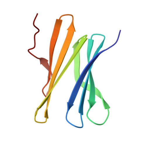
    Controlled protein assembly is an enabling technology, in particular, for biomaterials fabrication. Here, we report protein recognition and assembly by a phosphate-containing macrocycle ( pctx ). We show that the C 3 -symmetric phosphocavitand is a versatile receptor for N-terminal residues or arginine but not lysine. Using atomic resolution X-ray diffraction data, we reveal the precise details of N-terminal complexation in the β-propeller protein Ralstonia solanacearum lectin (RSL). In some cocrystal structures, a tetrahedral cluster of the phosphocavitand occupies one end of the β-propeller fold, providing a node for protein assembly. The macrocycle cluster is compatible with different types of precipitants, a broad pH range, and zinc complexation. We demonstrate system control with an arginine-enriched RSL that alters the overall assembly due to selective arginine complexation by pctx . A lysozyme- pctx cocrystal structure also demonstrates arginine complexation by the macrocycle. An alternative macrocycle cluster occurs with an engineered RSL bearing an extended N-terminus. In this structure, involving zinc ligation at the N-terminus, the macrocycle forms trimeric clusters and four such clusters form cage-like substructures within the tetrahedral protein framework. Thus, N-terminal complexation in combination with phosphocavitand self-assembly provides new routes to protein crystal engineering.


  • Organizational Affiliation
    • School of Biological and Chemical Sciences, University of Galway, Galway H91 TK33, Ireland.

Macromolecule Content 

  • Total Structure Weight: 11.45 kDa 
  • Atom Count: 926 
  • Modeled Residue Count: 90 
  • Deposited Residue Count: 90 
  • Unique protein chains: 1

Macromolecules

Find similar proteins by:|  3D Structure
Entity ID: 1
MoleculeChains  Sequence LengthOrganismDetailsImage
Fucose-binding lectin protein90Ralstonia solanacearumMutation(s): 3 
Gene Names: E7Z57_08365RSP795_21825RSP822_19650RUN39_v1_50103
UniProt
Find proteins for A0AAP7ZHZ3 (Ralstonia solanacearum K60)
Explore A0AAP7ZHZ3 
Go to UniProtKB:  A0AAP7ZHZ3
Entity Groups
Sequence Clusters30% Identity50% Identity70% Identity90% Identity95% Identity100% Identity
UniProt GroupA0AAP7ZHZ3
Sequence Annotations
Expand
Reference Sequence

Experimental Data & Validation

Experimental Data

  • Method: X-RAY DIFFRACTION
  • Resolution: 1.49 Å
  • R-Value Free:  0.203 (Depositor), 0.201 (DCC) 
  • R-Value Work:  0.172 (Depositor), 0.171 (DCC) 
  • R-Value Observed: 0.173 (Depositor) 
Space Group: H 3
Unit Cell:
Length ( Å )Angle ( ˚ )
a = 43.569α = 90
b = 43.569β = 90
c = 122.997γ = 120
Software Package:
Software NamePurpose
PHENIXrefinement
XDSdata reduction
Aimlessdata scaling
PHASERphasing
autoPROCdata processing

Structure Validation

View Full Validation Report


Ligand Structure Quality Assessment 


Entry History 

& Funding Information

Deposition Data


Funding OrganizationLocationGrant Number
Science Foundation IrelandIreland12/RC/2275_P2

Revision History  (Full details and data files)

  • Version 1.0: 2025-08-20
    Type: Initial release