9HN8 | pdb_00009hn8

Apo Structure of Truncated 1-deoxy-D-xylulose 5-phosphate synthase (DXPS) from Mycobacterium tuberculosis


Experimental Data Snapshot

  • Method: X-RAY DIFFRACTION
  • Resolution: 2.65 Å
  • R-Value Free: 
    0.273 (Depositor), 0.273 (DCC) 
  • R-Value Work: 
    0.247 (Depositor), 0.247 (DCC) 
  • R-Value Observed: 
    0.249 (Depositor) 

Starting Model: experimental
View more details

wwPDB Validation 3D Report Full Report

Validation slider image for 9HN8

This is version 1.0 of the entry. See complete history

Literature

Apo structure of Mycobacterium tuberculosis 1-deoxy-d-xylulose 5-phosphate synthase DXPS: Dynamics and implications for inhibitor design.

Gawriljuk, V.O.Alhayek, A.Hirsch, A.K.H.Groves, M.R.

(2025) Biochem Biophys Res Commun 747: 151246-151246

  • DOI: https://doi.org/10.1016/j.bbrc.2024.151246
  • Primary Citation Related Structures: 
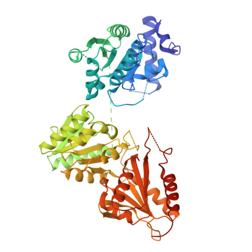
    9HN8

  • PubMed Abstract: 

    The enzyme 1-deoxy-d-xylulose-5-phosphate synthase (DXPS) catalyses the first step of the MEP pathway, a key process for isoprenoid biosynthesis in bacteria that is absent in humans, making it a promising drug target. We present the structure of Mycobacterium tuberculosis DXPS in its apo form, obtained through a soaking method that removes thiamine diphosphate (ThDP) and metals from pre-formed crystals. The apo structure had three regions with absence of electron density near the active site that are unique to the apo form of the enzyme. Comparisons with other homologous DXPS structures highlight a similar dynamic response to cofactor absence. Despite the increased flexibility, key residues for the activity and ThDP binding retain their positions, preserving the structural integrity of the catalytic core. These findings demonstrate the critical role of ThDP in maintaining DXPS stability and suggest that dynamic structural changes in the apo state may influence inhibitor binding targeting the cofactor site.


  • Organizational Affiliation
    • Chemical and Pharmaceutical Biology, Groningen Research Institute of Pharmacy, University of Groningen, Antonius Deusinglaan 1, 9713 AV, Groningen, the Netherlands.

Macromolecule Content 

  • Total Structure Weight: 127.25 kDa 
  • Atom Count: 7,205 
  • Modeled Residue Count: 954 
  • Deposited Residue Count: 1,208 
  • Unique protein chains: 1

Macromolecules

Find similar proteins by:|  3D Structure
Entity ID: 1
MoleculeChains  Sequence LengthOrganismDetailsImage
1-deoxy-D-xylulose-5-phosphate synthase
A, B
604Mycobacterium tuberculosis H37RvMutation(s): 0 
Gene Names: dxsRv2682cMTCY05A6.03c
EC: 2.2.1.7
UniProt
Find proteins for P9WNS3 (Mycobacterium tuberculosis (strain ATCC 25618 / H37Rv))
Explore P9WNS3 
Go to UniProtKB:  P9WNS3
Entity Groups
Sequence Clusters30% Identity50% Identity70% Identity90% Identity95% Identity100% Identity
UniProt GroupP9WNS3
Sequence Annotations
Expand
Reference Sequence

Experimental Data & Validation

Experimental Data

  • Method: X-RAY DIFFRACTION
  • Resolution: 2.65 Å
  • R-Value Free:  0.273 (Depositor), 0.273 (DCC) 
  • R-Value Work:  0.247 (Depositor), 0.247 (DCC) 
  • R-Value Observed: 0.249 (Depositor) 
Space Group: P 1 21 1
Unit Cell:
Length ( Å )Angle ( ˚ )
a = 67.18α = 90
b = 126.772β = 107.7
c = 80.553γ = 90
Software Package:
Software NamePurpose
PHENIXrefinement
XDSdata reduction
Aimlessdata scaling
PHASERphasing

Structure Validation

View Full Validation Report


Entry History 

& Funding Information

Deposition Data


Funding OrganizationLocationGrant Number
H2020 Marie Curie Actions of the European CommissionEuropean Union860816

Revision History  (Full details and data files)

  • Version 1.0: 2025-01-22
    Type: Initial release