9HLC | pdb_00009hlc

TRPML1 in complex with compound 11


Experimental Data Snapshot

  • Method: ELECTRON MICROSCOPY
  • Resolution: 2.10 Å
  • Aggregation State: PARTICLE 
  • Reconstruction Method: SINGLE PARTICLE 

Starting Model: experimental
View more details

wwPDB Validation 3D Report Full Report

Validation slider image for 9HLC

This is version 1.1 of the entry. See complete history. 

Literature

High throughput cryo-EM provides structural understanding for modulators of the lysosomal ion channel TRPML1.

Reeks, J., Mahajan, P., Clark, M., Cowan, S.R., Di Daniel, E., Earl, C.P., Fisher, S., Holvey, R.S., Jackson, S.M., Lloyd-Evans, E., Morgillo, C.M., Mortenson, P.N., O'Reilly, M., Richardson, C.J., Schopf, P., Tams, D.M., Waller-Evans, H., Ward, S.E., Whibley, S., Williams, P.A., Johnson, C.N.

(2025) Structure 33: 1374-1385.e7

  • DOI: https://doi.org/10.1016/j.str.2025.05.014
  • Primary Citation Related Structures: 
    9HJ6, 9HJ8, 9HL3, 9HL4, 9HL6, 9HL8, 9HLA, 9HLB, 9HLC, 9HLD

  • PubMed Abstract: 

    Access to high-resolution structural data for protein-ligand complexes is a prerequisite for structure-based medicinal chemistry, where the ability to iterate cycles of design-structure-redesign is highly desirable. For proteins refractory to X-ray crystallography, such as integral membrane proteins, enablement of high throughput structure determination by cryoelectron microscopy (cryo-EM) has the potential to be transformational for structure-based design. We have applied such an approach to the lysosomal ion channel transient receptor potential mucolipin 1 (TRPML1) in complex with ten chemically diverse modulators, both agonists and antagonists. The resulting depth of high-resolution structural data generated provides important insights into protein-ligand structure-function relationships, including mechanistic understanding of ligand-induced channel pore opening and closing. Moreover, the knowledge gained has the potential to support iterative design cycles toward improved modulators of this important biological target.


  • Organizational Affiliation: 
    • Astex Pharmaceuticals, 436 Cambridge Science Park, Milton Road, Cambridge CB4 0QA, UK.

Macromolecule Content 

  • Total Structure Weight: 291.2 kDa 
  • Atom Count: 15,756 
  • Modeled Residue Count: 1,832 
  • Deposited Residue Count: 2,512 
  • Unique protein chains: 1

Macromolecules

Find similar proteins by:|  3D Structure
Entity ID: 1
MoleculeChains  Sequence LengthOrganismDetailsImage
Mucolipin-1
A, B, C, D
628Homo sapiensMutation(s): 0 
Gene Names: MCOLN1, ML4, TRPML1, MSTP080
UniProt & NIH Common Fund Data Resources
Find proteins for Q9GZU1 (Homo sapiens)
Explore Q9GZU1 
Go to UniProtKB:  Q9GZU1
PHAROS:  Q9GZU1
GTEx:  ENSG00000090674 
Entity Groups
Sequence Clusters30% Identity50% Identity70% Identity90% Identity95% Identity100% Identity
UniProt GroupQ9GZU1
Glycosylation
Glycosylation Sites: 1Go to GlyGen: Q9GZU1-1
Sequence Annotations
Expand
Reference Sequence

Oligosaccharides

Help  
Entity ID: 2
MoleculeChains Length2D Diagram GlycosylationD Interactions
2-acetamido-2-deoxy-beta-D-glucopyranose-(1-4)-2-acetamido-2-deoxy-beta-D-glucopyranose
E, F, G, H
2N-Glycosylation
Glycosylation Resources
GlyTouCan: G42666HT
GlyCosmos: G42666HT
GlyGen: G42666HT

Small Molecules

Ligands 5 Unique
IDChains Name / Formula / InChI Key2D Diagram3D Interactions
EUJ

Query on EUJ


Download:Ideal Coordinates CCD File
BA [auth C],
JA [auth D],
L [auth A],
U [auth B]
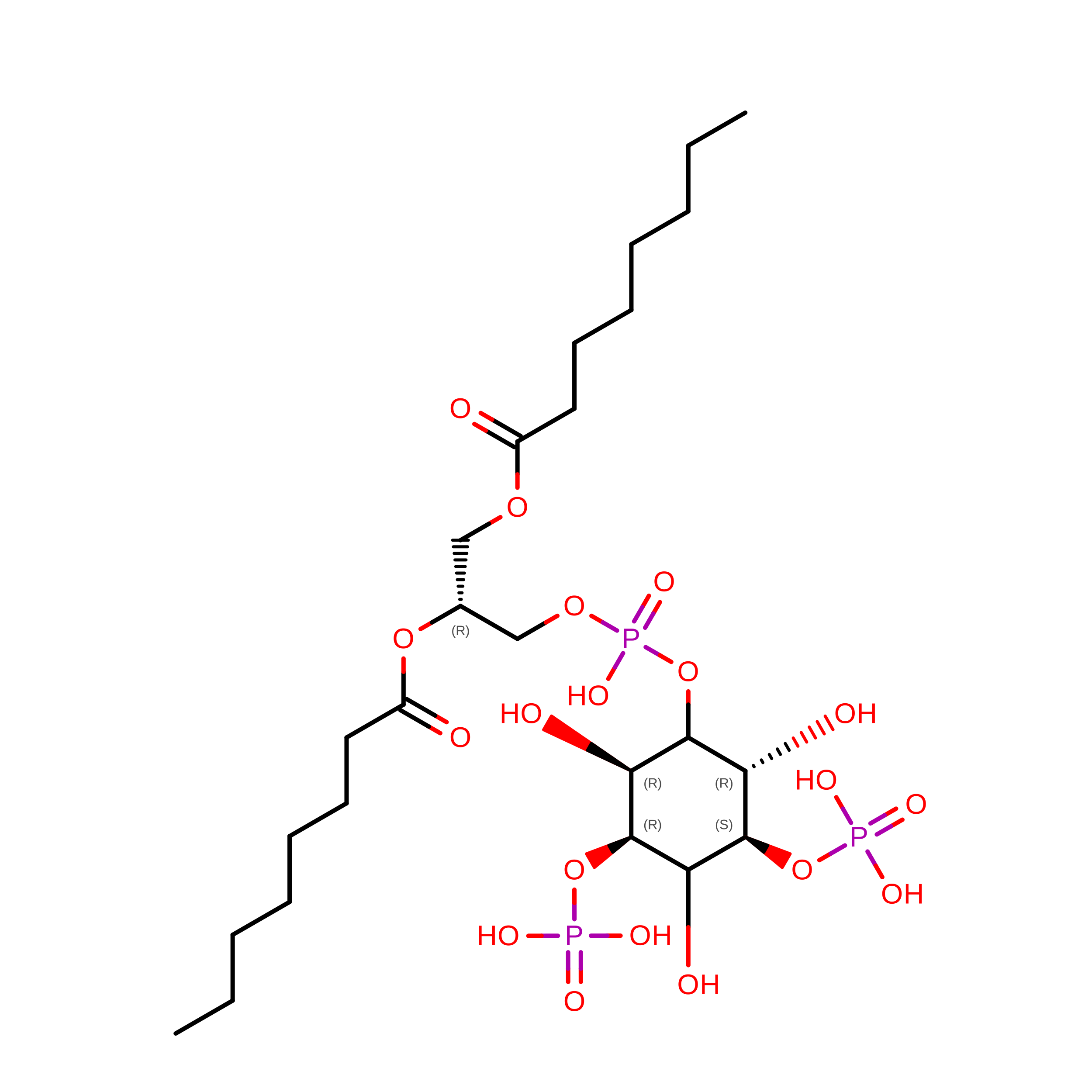
(2R)-3-{[(S)-hydroxy{[(1S,2R,3R,4S,5S,6R)-2,4,6-trihydroxy-3,5-bis(phosphonooxy)cyclohexyl]oxy}phosphoryl]oxy}propane-1,2-diyl dioctanoate
C25 H49 O19 P3
QXHVLVSULWMTCV-DICZBTHZSA-N
A1IV5
(Subject of Investigation/LOI)

Query on A1IV5


Download:Ideal Coordinates CCD File
CA [auth C],
DA [auth D],
M [auth A],
V [auth B]
(8R,9S,13S,14S,17S)-3-methoxy-13-methyl-6,7,8,9,11,12,14,15,16,17-decahydrocyclopenta[a]phenanthren-17-ol
C19 H26 O2
ULAADVBNYHGIBP-GFEQUFNTSA-N
8K6

Query on 8K6


Download:Ideal Coordinates CCD File
EA [auth D],
N [auth A],
P [auth B],
W [auth C]
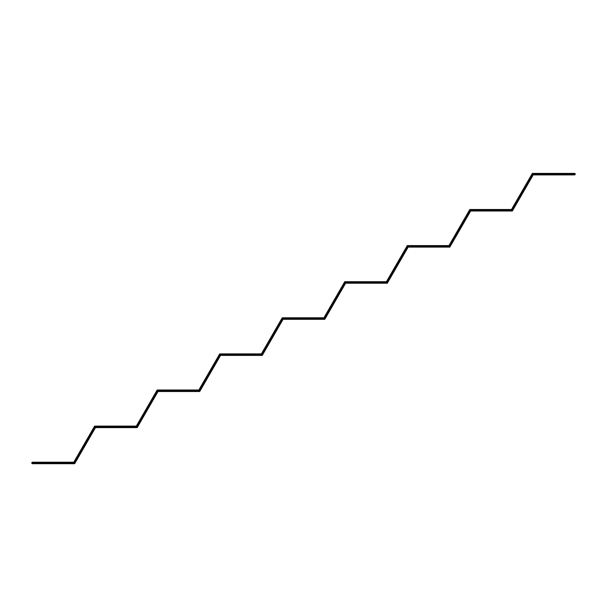
Octadecane
C18 H38
RZJRJXONCZWCBN-UHFFFAOYSA-N
D12

Query on D12


Download:Ideal Coordinates CCD File
AA [auth C]
FA [auth D]
IA [auth D]
K [auth A]
O [auth A]
AA [auth C],
FA [auth D],
IA [auth D],
K [auth A],
O [auth A],
Q [auth B],
T [auth B],
X [auth C]
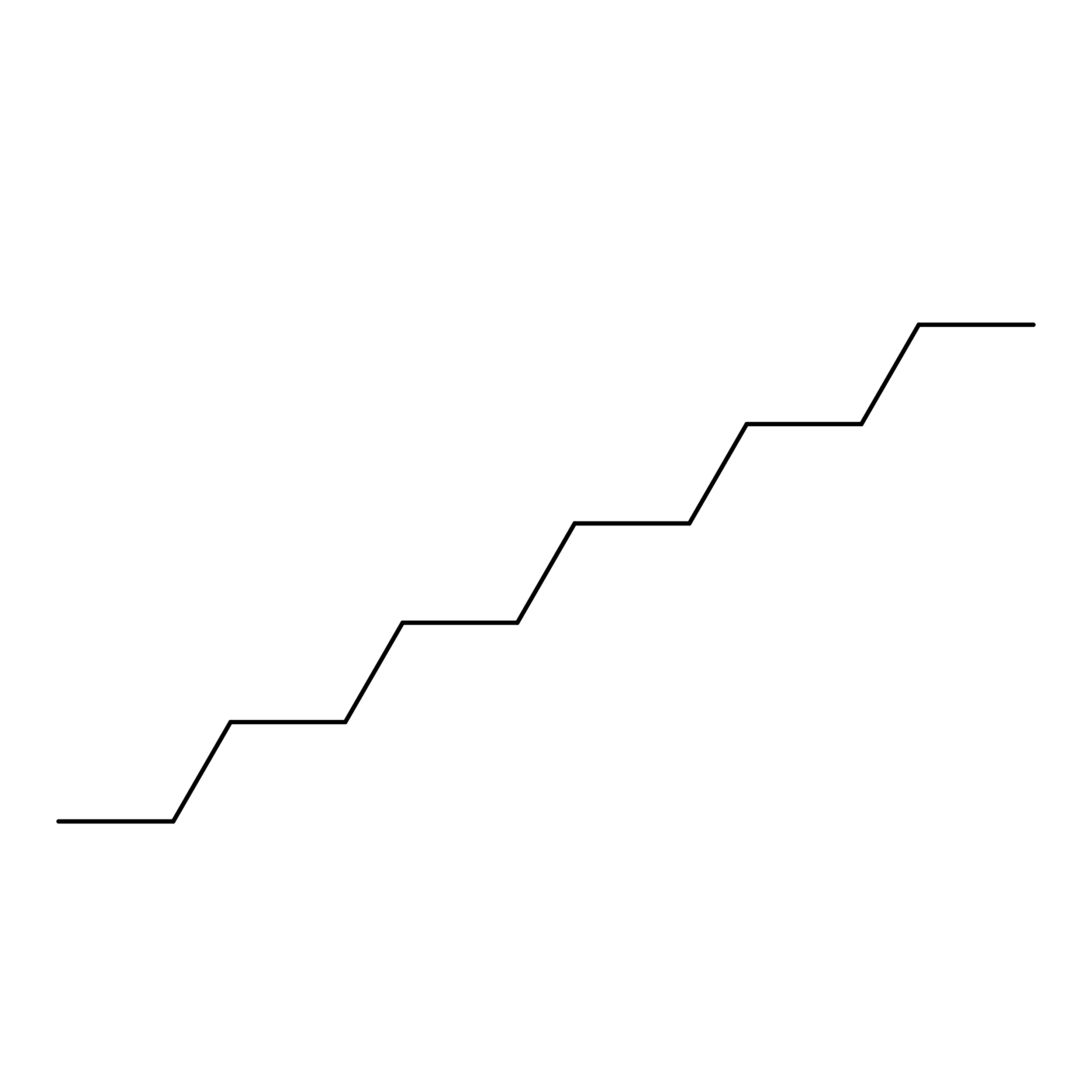
DODECANE
C12 H26
SNRUBQQJIBEYMU-UHFFFAOYSA-N
OCT

Query on OCT


Download:Ideal Coordinates CCD File
GA [auth D]
HA [auth D]
I [auth A]
J [auth A]
R [auth B]
GA [auth D],
HA [auth D],
I [auth A],
J [auth A],
R [auth B],
S [auth B],
Y [auth C],
Z [auth C]
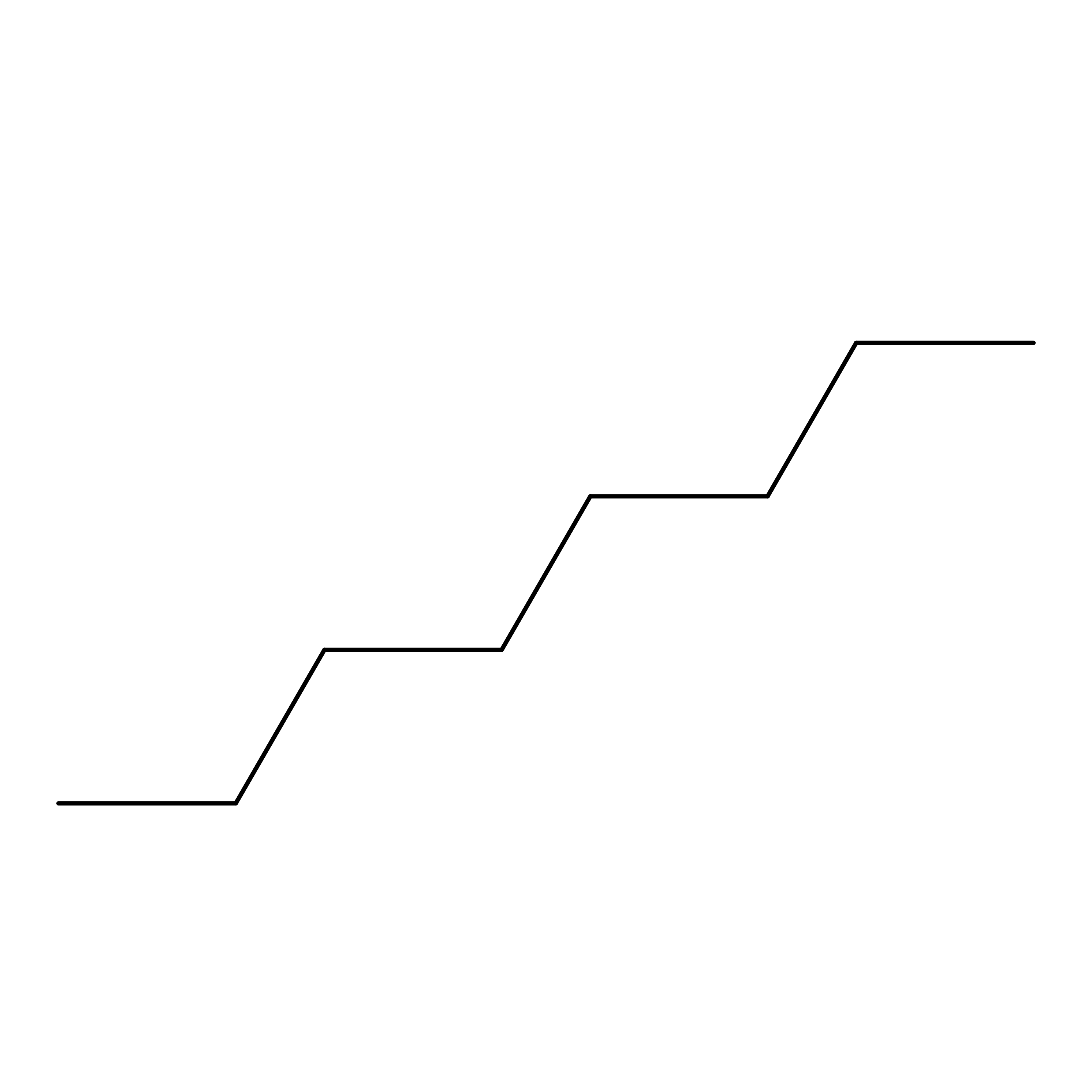
N-OCTANE
C8 H18
TVMXDCGIABBOFY-UHFFFAOYSA-N

Experimental Data & Validation

Experimental Data

  • Method: ELECTRON MICROSCOPY
  • Resolution: 2.10 Å
  • Aggregation State: PARTICLE 
  • Reconstruction Method: SINGLE PARTICLE 
EM Software:
TaskSoftware PackageVersion
RECONSTRUCTIONcryoSPARC4
MODEL REFINEMENTPHENIX1.21rc1-5101

Structure Validation

View Full Validation Report


Entry History 

& Funding Information

Revision History  (Full details and data files)

  • Version 1.0: 2025-06-04
    Type: Initial release
  • Version 1.1: 2025-12-17
    Changes: Data collection, Database references