9HHY | pdb_00009hhy

Crystal Structure of the Coxiella burnetii 2-methylisocitrate lyase Bound to Inhibitor Isocitric Acid


Experimental Data Snapshot

  • Method: X-RAY DIFFRACTION
  • Resolution: 2.33 Å
  • R-Value Free: 
    0.265 (Depositor), 0.265 (DCC) 
  • R-Value Work: 
    0.214 (Depositor), 0.214 (DCC) 
  • R-Value Observed: 
    0.217 (Depositor) 

Starting Model: in silico
View more details

wwPDB Validation 3D Report Full Report

Validation slider image for 9HHY

Ligand Structure Quality Assessment 


This is version 1.0 of the entry. See complete history

Literature

Structure and catalytic mechanism of methylisocitrate lyase, a potential drug target against Coxiella burnetii.

Stuart, W.S.Jenkins, C.H.Ireland, P.M.Isupov, M.N.Norville, I.H.Harmer, N.J.

(2025) J Biological Chem 301: 108517-108517

  • DOI: https://doi.org/10.1016/j.jbc.2025.108517
  • Primary Citation Related Structures: 
    9HGK, 9HGO, 9HGQ, 9HHS, 9HHY, 9HRA

  • PubMed Abstract: 

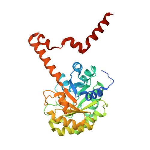
    We present a comprehensive investigation into the catalytic mechanism of methylisocitrate lyase, a potential drug target candidate against the zoonotic pathogen Coxiella burnetii, the causative agent of Q fever and a federal select agent. Current treatment regimens are prolonged, often with incomplete clearance of the pathogen. We utilized a structure-based bioinformatics pipeline to identify methylisocitrate lyase as a candidate therapeutic target against C. burnetii from a list of essential genes. WT C. burnetii methylisocitrate lyase has a k cat of 13.8 s -1 (compared to 105 s -1 for Salmonella enterica), and isocitrate inhibits with a K I of 11 mM. We have determined the previously uncharacterized substrate-bound structure of this enzyme family, alongside product and inhibitor-bound structures. These structures of WT enzyme reveal that in the active state the catalytic C118 is positioned 2.98 Å from O5 of methylisocitrate and Arg152 moves toward the substrate relative to the inhibitor bound structure. Analysis of structure-based mutants reveals that Arg152 and Glu110 are both essential for catalysis. We suggest that Arg152 acts as the catalytic base that initiates the methylisocitrate lyase reaction. These results deepen our understanding of the catalytic mechanism of methylisocitrate lyase and could aid the development of new therapeutics against C. burnetii.


  • Organizational Affiliation
    • Living Systems Institute, University of Exeter, Exeter, UK; Department of Biosciences, University of Exeter, Exeter, UK.

Macromolecule Content 

  • Total Structure Weight: 417.6 kDa 
  • Atom Count: 27,733 
  • Modeled Residue Count: 3,456 
  • Deposited Residue Count: 3,744 
  • Unique protein chains: 1

Macromolecules

Find similar proteins by:|  3D Structure
Entity ID: 1
MoleculeChains  Sequence LengthOrganismDetailsImage
2-methylisocitrate lyase
A, B, C, D, E
A, B, C, D, E, F, G, H, I, J, K, L
312Coxiella burnetiiMutation(s): 0 
Gene Names: prpBCBU_0771
EC: 4.1.3.30
UniProt
Find proteins for Q83DG5 (Coxiella burnetii (strain RSA 493 / Nine Mile phase I))
Explore Q83DG5 
Go to UniProtKB:  Q83DG5
Entity Groups
Sequence Clusters30% Identity50% Identity70% Identity90% Identity95% Identity100% Identity
UniProt GroupQ83DG5
Sequence Annotations
Expand
Reference Sequence

Small Molecules

Ligands 6 Unique
IDChains Name / Formula / InChI Key2D Diagram3D Interactions
ICT
(Subject of Investigation/LOI)

Query on ICT


Download:Ideal Coordinates CCD File
DA [auth D]
EB [auth H]
M [auth A]
MA [auth E]
MB [auth I]
DA [auth D],
EB [auth H],
M [auth A],
MA [auth E],
MB [auth I],
QA [auth F],
QB [auth J],
R [auth B],
VB [auth K],
WA [auth G],
Y [auth C],
ZB [auth L]
ISOCITRIC ACID
C6 H8 O7
ODBLHEXUDAPZAU-ZAFYKAAXSA-N
PGE

Query on PGE


Download:Ideal Coordinates CCD File
SB [auth J]TRIETHYLENE GLYCOL
C6 H14 O4
ZIBGPFATKBEMQZ-UHFFFAOYSA-N
PEG

Query on PEG


Download:Ideal Coordinates CCD File
NA [auth E]DI(HYDROXYETHYL)ETHER
C4 H10 O3
MTHSVFCYNBDYFN-UHFFFAOYSA-N
EDO

Query on EDO


Download:Ideal Coordinates CCD File
AB [auth G]
AC [auth L]
BB [auth G]
BC [auth L]
CA [auth D]
AB [auth G],
AC [auth L],
BB [auth G],
BC [auth L],
CA [auth D],
EA [auth D],
FA [auth D],
FB [auth H],
GA [auth D],
GB [auth H],
HA [auth D],
HB [auth H],
IA [auth D],
IB [auth H],
JA [auth D],
JB [auth H],
N [auth A],
NB [auth I],
O [auth A],
RA [auth F],
RB [auth J],
S [auth B],
SA [auth F],
T [auth B],
TA [auth F],
U [auth B],
V [auth B],
WB [auth K],
XA [auth G],
YA [auth G],
Z [auth C],
ZA [auth G]
1,2-ETHANEDIOL
C2 H6 O2
LYCAIKOWRPUZTN-UHFFFAOYSA-N
CL

Query on CL


Download:Ideal Coordinates CCD File
BA [auth C]
DB [auth G]
DC [auth L]
EC [auth L]
LA [auth D]
BA [auth C],
DB [auth G],
DC [auth L],
EC [auth L],
LA [auth D],
LB [auth H],
PA [auth E],
PB [auth I],
Q [auth A],
UB [auth J],
VA [auth F],
X [auth B],
YB [auth K]
CHLORIDE ION
Cl
VEXZGXHMUGYJMC-UHFFFAOYSA-M
MG
(Subject of Investigation/LOI)

Query on MG


Download:Ideal Coordinates CCD File
AA [auth C]
CB [auth G]
CC [auth L]
KA [auth D]
KB [auth H]
AA [auth C],
CB [auth G],
CC [auth L],
KA [auth D],
KB [auth H],
OA [auth E],
OB [auth I],
P [auth A],
TB [auth J],
UA [auth F],
W [auth B],
XB [auth K]
MAGNESIUM ION
Mg
JLVVSXFLKOJNIY-UHFFFAOYSA-N

Experimental Data & Validation

Experimental Data

  • Method: X-RAY DIFFRACTION
  • Resolution: 2.33 Å
  • R-Value Free:  0.265 (Depositor), 0.265 (DCC) 
  • R-Value Work:  0.214 (Depositor), 0.214 (DCC) 
  • R-Value Observed: 0.217 (Depositor) 
Space Group: P 1 21 1
Unit Cell:
Length ( Å )Angle ( ˚ )
a = 93.785α = 90
b = 115.55β = 92.34
c = 195.241γ = 90
Software Package:
Software NamePurpose
REFMACrefinement
xia2data reduction
DIALSdata reduction
Aimlessdata scaling
PHASERphasing

Structure Validation

View Full Validation Report


Ligand Structure Quality Assessment 


Entry History 

& Funding Information

Deposition Data


Funding OrganizationLocationGrant Number
Biotechnology and Biological Sciences Research Council (BBSRC)United KingdomBB/M009122/1

Revision History  (Full details and data files)

  • Version 1.0: 2025-08-13
    Type: Initial release