9HG1 | pdb_00009hg1

Crystal structure of M. smegmatis GMP reductase in complex with GMP and ATP.


Experimental Data Snapshot

  • Method: X-RAY DIFFRACTION
  • Resolution: 2.30 Å
  • R-Value Free: 
    0.230 (Depositor), 0.229 (DCC) 
  • R-Value Work: 
    0.200 (Depositor), 0.200 (DCC) 
  • R-Value Observed: 
    0.201 (Depositor) 

Starting Model: experimental
View more details

wwPDB Validation 3D Report Full Report

Validation slider image for 9HG1

Ligand Structure Quality Assessment 


This is version 1.1 of the entry. See complete history

Literature

Structural basis for allosteric regulation of mycobacterial guanosine 5 ́-monophosphate reductase by ATP and GTP.

Dolezal, M.Knejzlik, Z.Kouba, T.Filimonenko, A.Svachova, H.Dedola, M.Klima, M.Pichova, I.

(2026) Nat Commun 

  • DOI: https://doi.org/10.1038/s41467-026-71657-9
  • Primary Citation Related Structures: 
    8RY0, 8RY1, 8RY3, 8RY4, 8RY5, 8RY6, 8RY7, 8RY8, 8RY9, 8RYA, 8RYB, 9HFZ, 9HG0, 9HG1, 9HG2, 9HG3

  • PubMed Abstract: 

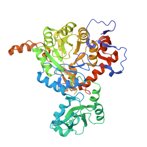
    Guanosine 5'-monophosphate reductase (GMPR) is a crucial enzyme in the purine salvage pathway that catalyses the NADPH-dependent conversion of GMP to IMP, thereby contributing to purine nucleotide homeostasis. Mycobacterium smegmatis GMPR (MsmGMPR) contains a regulatory cystathionine β-synthase (CBS) domain, which mediates allosteric modulation by ATP and GTP. However, MsmGMPR exhibits an atypical tertiary structure that is incompatible with the acknowledged regulatory mechanisms of IMPDH/GMPR family enzymes. Here, we combine X-ray crystallography, cryogenic electron microscopy, and biochemical binding assays to elucidate the molecular basis of MsmGMPR regulation by ATP and GTP. We show that ATP stabilises a compressed conformation that inhibits the enzyme by restricting access to the active site and preventing NADPH binding. In contrast, GTP counteracts ATP binding, promoting an active conformation that enables catalysis. Our results provide insight into how MsmGMPR senses and responds to the purine nucleotide balance, revealing a distinct utilisation of the CBS domain compared with its typical role in IMPDH/GMPR enzymes.


  • Organizational Affiliation
    • Institute of Organic Chemistry and Biochemistry of the Czech Academy of Sciences, Prague, Czech Republic.

Macromolecule Content 

  • Total Structure Weight: 842.45 kDa 
  • Atom Count: 57,130 
  • Modeled Residue Count: 7,568 
  • Deposited Residue Count: 7,936 
  • Unique protein chains: 1

Macromolecules

Find similar proteins by:|  3D Structure
Entity ID: 1
MoleculeChains  Sequence LengthOrganismDetailsImage
GMP reductase
A, B, C, D, E
A, B, C, D, E, F, G, H, I, J, K, L, M, N, O, P
496Mycolicibacterium smegmatisMutation(s): 0 
Gene Names: guaB1MSMEG_3634MSMEI_3548
EC: 1.7.1.7
UniProt
Find proteins for A0QYE8 (Mycolicibacterium smegmatis (strain ATCC 700084 / mc(2)155))
Explore A0QYE8 
Go to UniProtKB:  A0QYE8
Entity Groups
Sequence Clusters30% Identity50% Identity70% Identity90% Identity95% Identity100% Identity
UniProt GroupA0QYE8
Sequence Annotations
Expand
Reference Sequence

Small Molecules

Ligands 2 Unique
IDChains Name / Formula / InChI Key2D Diagram3D Interactions
ATP
(Subject of Investigation/LOI)

Query on ATP


Download:Ideal Coordinates CCD File
AA [auth F]
CA [auth G]
EA [auth H]
GA [auth I]
IA [auth J]
AA [auth F],
CA [auth G],
EA [auth H],
GA [auth I],
IA [auth J],
KA [auth K],
NA [auth L],
PA [auth M],
Q [auth A],
RA [auth N],
S [auth B],
TA [auth O],
U [auth C],
VA [auth P],
W [auth D],
Y [auth E]
ADENOSINE-5'-TRIPHOSPHATE
C10 H16 N5 O13 P3
ZKHQWZAMYRWXGA-KQYNXXCUSA-N
5GP
(Subject of Investigation/LOI)

Query on 5GP


Download:Ideal Coordinates CCD File
BA [auth F]
DA [auth G]
FA [auth H]
HA [auth I]
JA [auth J]
BA [auth F],
DA [auth G],
FA [auth H],
HA [auth I],
JA [auth J],
LA [auth K],
MA [auth L],
OA [auth M],
QA [auth N],
R [auth A],
SA [auth O],
T [auth B],
UA [auth P],
V [auth C],
X [auth D],
Z [auth E]
GUANOSINE-5'-MONOPHOSPHATE
C10 H14 N5 O8 P
RQFCJASXJCIDSX-UUOKFMHZSA-N

Experimental Data & Validation

Experimental Data

  • Method: X-RAY DIFFRACTION
  • Resolution: 2.30 Å
  • R-Value Free:  0.230 (Depositor), 0.229 (DCC) 
  • R-Value Work:  0.200 (Depositor), 0.200 (DCC) 
  • R-Value Observed: 0.201 (Depositor) 
Space Group: P 1
Unit Cell:
Length ( Å )Angle ( ˚ )
a = 112.57α = 82.7
b = 113.6β = 77.236
c = 178.06γ = 66.749
Software Package:
Software NamePurpose
XDSdata reduction
XSCALEdata scaling
PHASERphasing
Cootmodel building
PHENIXrefinement

Structure Validation

View Full Validation Report


Ligand Structure Quality Assessment 


Entry History 

& Funding Information

Deposition Data


Funding OrganizationLocationGrant Number
European Union (EU)European UnionLX22NPO5103

Revision History  (Full details and data files)

  • Version 1.0: 2025-08-27
    Type: Initial release
  • Version 1.1: 2026-04-22
    Changes: Database references