9H6L | pdb_00009h6l

Human B4GALNT1 in Complex with UDP


Experimental Data Snapshot

  • Method: X-RAY DIFFRACTION
  • Resolution: 2.67 Å
  • R-Value Free: 
    0.233 (Depositor), 0.233 (DCC) 
  • R-Value Work: 
    0.191 (Depositor), 0.191 (DCC) 

Starting Model: in silico
View more details

wwPDB Validation 3D Report Full Report

Validation slider image for 9H6L

Ligand Structure Quality Assessment 


This is version 1.0 of the entry. See complete history

Literature

Conformational dynamics and membrane insertion mechanism of B4GALNT1 in ganglioside synthesis.

Welland, J.W.J.Barrow, H.G.Stansfeld, P.J.Deane, J.E.

(2025) Nat Commun 16: 5442-5442

  • DOI: https://doi.org/10.1038/s41467-025-60593-9
  • Primary Citation Related Structures: 
    9H6J, 9H6K, 9H6L

  • PubMed Abstract: 

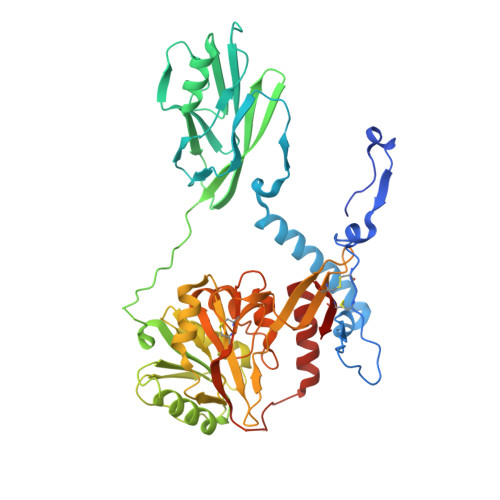
    Glycosphingolipids (GSLs) are crucial membrane components involved in essential cellular pathways. Complex GSLs, known as gangliosides, are synthesised by glycosyltransferase enzymes and imbalances in GSL metabolism cause severe neurological diseases. B4GALNT1 synthesises the precursors to the major brain gangliosides. Loss of B4GALNT1 function causes hereditary spastic paraplegia, while its overexpression is linked to cancers including childhood neuroblastoma. Here, we present crystal structures of the human homodimeric B4GALNT1 enzyme demonstrating dynamic remodelling of the substrate binding site during catalysis. We show that processing of lipid substrates by B4GALNT1 is severely compromised when surface loops flanking the active site are mutated from hydrophobic residues to polar. Molecular dynamics simulations support that these loops can insert into the lipid bilayer explaining how B4GALNT1 accesses and processes lipid substrates. By combining structure prediction and molecular simulations we propose that this mechanism of dynamic membrane insertion is exploited by other, structurally distinct GSL synthesising enzymes.


  • Organizational Affiliation
    • Cambridge Institute for Medical Research, Department of Clinical Neuroscience, University of Cambridge, Cambridge, UK.

Macromolecule Content 

  • Total Structure Weight: 113.42 kDa 
  • Atom Count: 7,349 
  • Modeled Residue Count: 918 
  • Deposited Residue Count: 998 
  • Unique protein chains: 1

Macromolecules

Find similar proteins by:|  3D Structure
Entity ID: 1
MoleculeChains  Sequence LengthOrganismDetailsImage
Beta-1,4 N-acetylgalactosaminyltransferase 1
A, B
499Homo sapiensMutation(s): 0 
Gene Names: B4GALNT1GALGTSIAT2
EC: 2.4.1.92
UniProt & NIH Common Fund Data Resources
Find proteins for Q00973 (Homo sapiens)
Explore Q00973 
Go to UniProtKB:  Q00973
PHAROS:  Q00973
GTEx:  ENSG00000135454 
Entity Groups
Sequence Clusters30% Identity50% Identity70% Identity90% Identity95% Identity100% Identity
UniProt GroupQ00973
Glycosylation
Glycosylation Sites: 1Go to GlyGen: Q00973-1
Sequence Annotations
Expand
Reference Sequence

Oligosaccharides

Help  
Entity ID: 2
MoleculeChains Length2D Diagram GlycosylationD Interactions
beta-D-mannopyranose-(1-4)-2-acetamido-2-deoxy-beta-D-glucopyranose-(1-4)-2-acetamido-2-deoxy-beta-D-glucopyranose
C, D
3N-Glycosylation
Glycosylation Resources
GlyTouCan: G15407YE
GlyCosmos: G15407YE
GlyGen: G15407YE

Small Molecules

Experimental Data & Validation

Experimental Data

  • Method: X-RAY DIFFRACTION
  • Resolution: 2.67 Å
  • R-Value Free:  0.233 (Depositor), 0.233 (DCC) 
  • R-Value Work:  0.191 (Depositor), 0.191 (DCC) 
Space Group: P 1 21 1
Unit Cell:
Length ( Å )Angle ( ˚ )
a = 60.183α = 90
b = 133.527β = 94.83
c = 86.922γ = 90
Software Package:
Software NamePurpose
REFMACrefinement
DIALSdata collection
Aimlessdata scaling
PHASERphasing
ISOLDEmodel building
Cootmodel building
DIALSdata reduction

Structure Validation

View Full Validation Report


Ligand Structure Quality Assessment 


Entry History 

& Funding Information

Deposition Data


Funding OrganizationLocationGrant Number
Wellcome TrustUnited Kingdom--

Revision History  (Full details and data files)

  • Version 1.0: 2025-07-16
    Type: Initial release