9H4F | pdb_00009h4f

Structure of Imine Reductase 361 from Micromonospora sp. mutant M125W/I127F/L179V/H250L


Experimental Data Snapshot

  • Method: X-RAY DIFFRACTION
  • Resolution: 2.77 Å
  • R-Value Free: 
    0.304 (Depositor), 0.306 (DCC) 
  • R-Value Work: 
    0.226 (Depositor), 0.233 (DCC) 
  • R-Value Observed: 
    0.229 (Depositor) 

Starting Model: experimental
View more details

wwPDB Validation 3D Report Full Report

Validation slider image for 9H4F

Ligand Structure Quality Assessment 


This is version 1.0 of the entry. See complete history

Literature

Engineered Biocatalyst for Enantioselective Hydrazone Reduction.

Hutton, A.E.Zhao, F.Ho, E.Domenech, J.Harawa, V.Brown, M.J.B.Grogan, G.Clayman, P.D.Turner, N.J.Green, A.P.

(2025) Angew Chem Int Ed Engl 64: e202424350-e202424350

  • DOI: https://doi.org/10.1002/anie.202424350
  • Primary Citation Related Structures: 
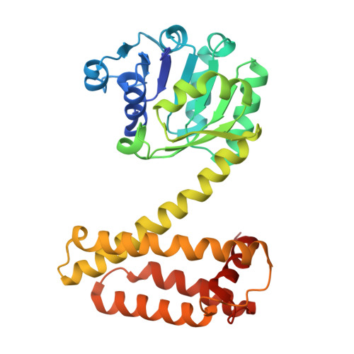
    9H4F

  • PubMed Abstract: 

    Enantioselective reduction of hydrazones provides a convergent and versatile route to synthesize hydrazine-containing motifs that are commonly found in pharmaceuticals and agrochemicals. However, current methods require the use of precious metals, costly chiral ligands, and/or forcing reaction conditions. Here, we report the development of a biocatalytic approach for enantioselective hydrazone reduction using engineered imine reductases. Following evaluation of an in-house panel of >400 IRED sequences, we identified a single IR361 I127F L179V variant that promotes reduction of Cbz-protected hydrazones. The introduction of additional two mutations via directed evolution afforded HRED1.1 that is 20-fold more active than the parent template and promotes reduction of a variety of protected hydrazones in high yields and selectivities (>99% e.e.), including in preparative scale biotransformations. Structural analysis of HRED1.1 provides insights into the origins of its unique hydrazone reductase activity. This study offers a powerful biocatalytic route to synthesize valuable chiral hydrazine products and further expands the impressive range of transformations accessible with engineered imine reductases.


  • Organizational Affiliation
    • Manchester Institute of Biotechnology and Department of Chemistry, University of Manchester, 131 Princess Street, Manchester, M1 7DN, UK.

Macromolecule Content 

  • Total Structure Weight: 63.97 kDa 
  • Atom Count: 3,785 
  • Modeled Residue Count: 579 
  • Deposited Residue Count: 594 
  • Unique protein chains: 1

Macromolecules

Find similar proteins by:|  3D Structure
Entity ID: 1
MoleculeChains  Sequence LengthOrganismDetailsImage
6-phosphogluconate dehydrogenase
A, B
297MicromonosporaMutation(s): 4 
Gene Names: BSA16_00815
UniProt
Find proteins for A0ACD6BA29 (Micromonospora sp. Rc5)
Explore A0ACD6BA29 
Go to UniProtKB:  A0ACD6BA29
Entity Groups
Sequence Clusters30% Identity50% Identity70% Identity90% Identity95% Identity100% Identity
UniProt GroupA0ACD6BA29
Sequence Annotations
Expand
Reference Sequence

Small Molecules

Ligands 1 Unique
IDChains Name / Formula / InChI Key2D Diagram3D Interactions
NAP
(Subject of Investigation/LOI)

Query on NAP


Download:Ideal Coordinates CCD File
C [auth A]NADP NICOTINAMIDE-ADENINE-DINUCLEOTIDE PHOSPHATE
C21 H28 N7 O17 P3
XJLXINKUBYWONI-NNYOXOHSSA-N

Experimental Data & Validation

Experimental Data

  • Method: X-RAY DIFFRACTION
  • Resolution: 2.77 Å
  • R-Value Free:  0.304 (Depositor), 0.306 (DCC) 
  • R-Value Work:  0.226 (Depositor), 0.233 (DCC) 
  • R-Value Observed: 0.229 (Depositor) 
Space Group: I 21 3
Unit Cell:
Length ( Å )Angle ( ˚ )
a = 184.268α = 90
b = 184.268β = 90
c = 184.268γ = 90
Software Package:
Software NamePurpose
REFMACrefinement
XDSdata reduction
SCALAdata scaling
MOLREPphasing

Structure Validation

View Full Validation Report


Ligand Structure Quality Assessment 


Entry History 

& Funding Information

Deposition Data


Funding OrganizationLocationGrant Number
Not funded--

Revision History  (Full details and data files)

  • Version 1.0: 2025-08-27
    Type: Initial release