


Structure of the SARS-CoV spike glycoprotein in complex with a homotrimeric Bicycle molecule
Drulyte, I., Pellegrino, S., Harman, M., Bezerra, G.A.To be published.
Experimental Data Snapshot
wwPDB Validation 3D Report Full Report

Entity ID: 1 | |||||
|---|---|---|---|---|---|
| Molecule | Chains | Sequence Length | Organism | Details | Image |
| Spike glycoprotein | 1,133 | Severe acute respiratory syndrome coronavirus 2 | Mutation(s): 6 Gene Names: S, 2 | 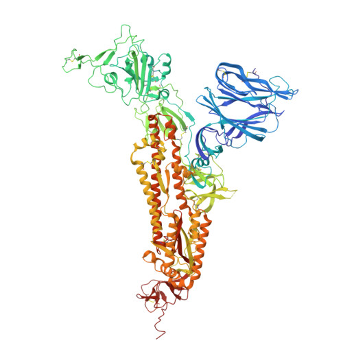 | |
UniProt | |||||
Entity Groups | |||||
| Sequence Clusters | 30% Identity50% Identity70% Identity90% Identity95% Identity100% Identity | ||||
| UniProt Group | P0DTC2 | ||||
Glycosylation | |||||
| Glycosylation Sites: 9 | Go to GlyGen: P0DTC2-1 | ||||
Sequence AnnotationsExpand | |||||
Reference Sequence | |||||
Entity ID: 2 | |||||
|---|---|---|---|---|---|
| Molecule | Chains | Sequence Length | Organism | Details | Image |
| Homotrimeric bicycle molecule | 16 | synthetic construct | Mutation(s): 0 |  | |
Entity Groups | |||||
| Sequence Clusters | 30% Identity50% Identity70% Identity90% Identity95% Identity100% Identity | ||||
Sequence AnnotationsExpand | |||||
Reference Sequence | |||||
Entity ID: 3 | |||||
|---|---|---|---|---|---|
| Molecule | Chains | Length | 2D Diagram | Glycosylation | D Interactions |
| 2-acetamido-2-deoxy-beta-D-glucopyranose-(1-4)-2-acetamido-2-deoxy-beta-D-glucopyranose | G [auth AA], H [auth AB], I [auth AC], J [auth BA], K [auth BB], G [auth AA], H [auth AB], I [auth AC], J [auth BA], K [auth BB], L [auth BC], M [auth CA], N [auth CB], O [auth CC] | 2 |  | N-Glycosylation | |
Glycosylation Resources | |||||
GlyTouCan: G42666HT GlyCosmos: G42666HT GlyGen: G42666HT | |||||
| Ligands 3 Unique | |||||
|---|---|---|---|---|---|
| ID | Chains | Name / Formula / InChI Key | 2D Diagram | 3D Interactions | |
| STE Download:Ideal Coordinates CCD File | FA [auth B], MA [auth C], W [auth A] | STEARIC ACID C18 H36 O2 QIQXTHQIDYTFRH-UHFFFAOYSA-N |  | ||
| KZ0 (Subject of Investigation/LOI) Download:Ideal Coordinates CCD File | NA [auth D], OA [auth E], PA [auth F] | 2,4,6-tris(chloromethyl)-1,3,5-triazine C6 H6 Cl3 N3 RICRAVHJCLFPFF-UHFFFAOYSA-N |  | ||
| NAG Download:Ideal Coordinates CCD File | AA [auth B] BA [auth B] CA [auth B] DA [auth B] EA [auth B] | 2-acetamido-2-deoxy-beta-D-glucopyranose C8 H15 N O6 OVRNDRQMDRJTHS-FMDGEEDCSA-N |  | ||
| Modified Residues 1 Unique | |||||
|---|---|---|---|---|---|
| ID | Chains | Type | Formula | 2D Diagram | Parent |
| 4J5 Query on 4J5 | D, E, F | L-PEPTIDE LINKING | C5 H13 N4 O2 |  | ARG |
| DAL Query on DAL | D, E, F | D-PEPTIDE LINKING | C3 H7 N O2 |  | -- |
| Task | Software Package | Version |
|---|---|---|
| MODEL REFINEMENT | PHENIX | 1.20.1_4487: |
| Funding Organization | Location | Grant Number |
|---|---|---|
| Other private | -- |


