9GTG | pdb_00009gtg

RIPK1 in complex with AZ"902


Experimental Data Snapshot

  • Method: X-RAY DIFFRACTION
  • Resolution: 2.25 Å
  • R-Value Free: 
    0.295 (Depositor), 0.293 (DCC) 
  • R-Value Work: 
    0.217 (Depositor), 0.227 (DCC) 
  • R-Value Observed: 
    0.221 (Depositor) 

Starting Model: experimental
View more details

wwPDB Validation 3D Report Full Report

Validation slider image for 9GTG

Ligand Structure Quality Assessment 


This is version 1.2 of the entry. See complete history

Literature

Discovery and Validation of a Novel Class of Necroptosis Inhibitors Targeting RIPK1.

Soday, L.Seripracharat, C.Gray, J.L.Luz, A.F.S.Howard, R.T.Singh, R.Burden, T.J.Bernardini, E.Mateus-Pinheiro, M.Petersen, J.Gunnarsson, A.Gunnarsson, J.Aagaard, A.Sjogren, T.Maslen, S.Bartlett, E.J.Iles, A.F.Smith, D.M.Scott, J.S.Skehel, M.Davis, A.M.Ressurreicao, A.S.Moreira, R.Rodrigues, C.M.P.Shenoy, A.R.Tate, E.W.

(2025) ACS Chem Biol 20: 1527-1543

  • DOI: https://doi.org/10.1021/acschembio.5c00112
  • Primary Citation Related Structures: 
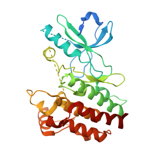
    9GTG, 9GTY

  • PubMed Abstract: 

    Necroptosis is a form of programmed cell death that, when dysregulated, is associated with cancer and inflammatory and neurodegenerative diseases. Here, starting from hits identified from a phenotypic high-throughput screen for inhibitors of necroptosis, we synthesized a library of compounds containing a 7-phenylquinoline motif and validated their anti-necroptotic activity in a novel live-cell assay. Based on these data, we designed an optimized photoaffinity probe for target engagement studies and through biochemical and cell-based assays established receptor-interacting kinase 1 (RIPK1) as the cellular target, with inhibition of necroptosis arising from the prevention of RIPK1 autophosphorylation and activation. X-ray crystallography and mass spectrometry revealed that these compounds bind at the hinge region of the active conformation of RIPK1, establishing them as type I kinase inhibitors. In addition, we demonstrated in vitro synergy with type III kinase inhibitors, such as necrostatin-1 and found that lead compounds protected mice against acute inflammation in necroptosis models in vivo . Overall, we present a novel pharmacophore for inhibition of human RIPK1, a key protein involved in necroptosis, and provide a photoaffinity probe to explore RIPK1 target engagement in cells.


  • Organizational Affiliation
    • Department of Chemistry, Molecular Sciences Research Hub, Imperial College London, London W12 0BZ, U.K.

Macromolecule Content 

  • Total Structure Weight: 72.59 kDa 
  • Atom Count: 4,225 
  • Modeled Residue Count: 518 
  • Deposited Residue Count: 626 
  • Unique protein chains: 1

Macromolecules

Find similar proteins by:|  3D Structure
Entity ID: 1
MoleculeChains  Sequence LengthOrganismDetailsImage
Receptor-interacting serine/threonine-protein kinase 1
A, B
313Homo sapiensMutation(s): 0 
Gene Names: RIPK1RIPRIP1
EC: 2.7.11.1
UniProt & NIH Common Fund Data Resources
Find proteins for Q13546 (Homo sapiens)
Explore Q13546 
Go to UniProtKB:  Q13546
PHAROS:  Q13546
GTEx:  ENSG00000137275 
Entity Groups
Sequence Clusters30% Identity50% Identity70% Identity90% Identity95% Identity100% Identity
UniProt GroupQ13546
Sequence Annotations
Expand
Reference Sequence

Experimental Data & Validation

Experimental Data

  • Method: X-RAY DIFFRACTION
  • Resolution: 2.25 Å
  • R-Value Free:  0.295 (Depositor), 0.293 (DCC) 
  • R-Value Work:  0.217 (Depositor), 0.227 (DCC) 
  • R-Value Observed: 0.221 (Depositor) 
Space Group: P 21 21 21
Unit Cell:
Length ( Å )Angle ( ˚ )
a = 47.084α = 90
b = 96.491β = 90
c = 129.002γ = 90
Software Package:
Software NamePurpose
REFMACrefinement
Aimlessdata scaling
MOLREPphasing

Structure Validation

View Full Validation Report


Ligand Structure Quality Assessment 


Entry History 

& Funding Information

Deposition Data

  • Released Date: 2025-07-02 
  • Deposition Author(s): Petersen, J.

Funding OrganizationLocationGrant Number
Not funded--

Revision History  (Full details and data files)

  • Version 1.0: 2025-07-02
    Type: Initial release
  • Version 1.1: 2025-07-16
    Changes: Source and taxonomy
  • Version 1.2: 2025-07-30
    Changes: Database references