


Cryo-electron microscopy structure of glucose/xylose isomerase from Streptomyces rubiginosus with magnesium ions in the active site
Slawek, J., Klonecka, A., Rawski, M., Kozak, M.To be published.
Experimental Data Snapshot
wwPDB Validation 3D Report Full Report

Entity ID: 1 | |||||
|---|---|---|---|---|---|
| Molecule | Chains | Sequence Length | Organism | Details | Image |
| Xylose isomerase | 388 | Streptomyces pseudogriseolus | Mutation(s): 0 EC: 5.3.1.5 | 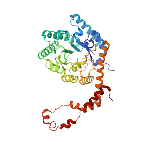 | |
UniProt | |||||
Entity Groups | |||||
| Sequence Clusters | 30% Identity50% Identity70% Identity90% Identity95% Identity100% Identity | ||||
| UniProt Group | P24300 | ||||
Sequence AnnotationsExpand | |||||
Reference Sequence | |||||
| Ligands 1 Unique | |||||
|---|---|---|---|---|---|
| ID | Chains | Name / Formula / InChI Key | 2D Diagram | 3D Interactions | |
| MG (Subject of Investigation/LOI) Download:Ideal Coordinates CCD File | E [auth A] F [auth A] G [auth B] H [auth B] I [auth C] | MAGNESIUM ION Mg JLVVSXFLKOJNIY-UHFFFAOYSA-N |  | ||
| Task | Software Package | Version |
|---|---|---|
| MODEL REFINEMENT | PHENIX | |
| RECONSTRUCTION | cryoSPARC |
| Funding Organization | Location | Grant Number |
|---|---|---|
| Polish National Science Centre | Poland | 2020/39/O/ST4/03465 |


