9GM3 | pdb_00009gm3

Crystal structure of the complex formed between the radical SAM protein ChlB and the leader region of its precursor substrate ChlA


Experimental Data Snapshot

  • Method: X-RAY DIFFRACTION
  • Resolution: 1.65 Å
  • R-Value Free: 
    0.213 (Depositor), 0.204 (DCC) 
  • R-Value Work: 
    0.193 (Depositor), 0.186 (DCC) 
  • R-Value Observed: 
    0.194 (Depositor) 

Starting Model: in silico
View more details

wwPDB Validation 3D Report Full Report

Validation slider image for 9GM3

Ligand Structure Quality Assessment 


This is version 1.0 of the entry. See complete history

Literature

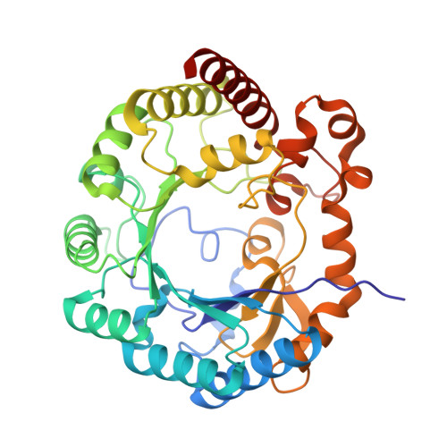
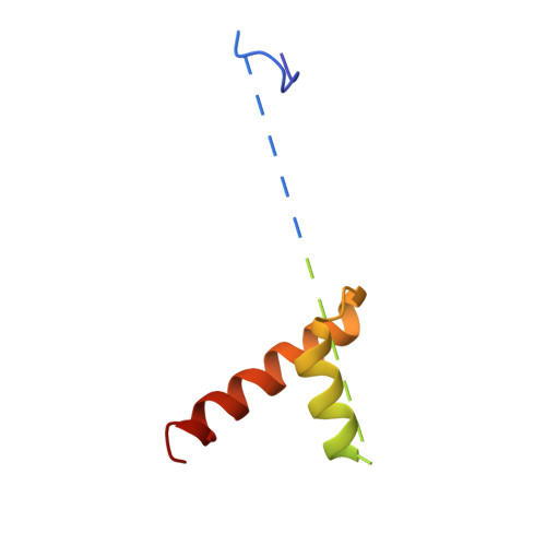
Peptide Recognition and Mechanism of the Radical S -Adenosyl-l-methionine Multiple Cyclophane Synthase ChlB.

Ruel, J.Nguyen, T.Q.N.Morishita, Y.Usclat, A.Martin, L.Amara, P.Kieffer-Jaquinod, S.Stefanoiu, M.C.de la Mora, E.Morinaka, B.I.Nicolet, Y.

(2025) J Am Chem Soc 147: 16850-16863

  • DOI: https://doi.org/10.1021/jacs.4c16004
  • Primary Citation Related Structures: 
    9GM3, 9GMC

  • PubMed Abstract: 

    Ribosomally synthesized and post-translationally modified peptides (RiPPs) represent a valuable class of natural products, often featuring macrocyclization, which enhances stability and rigidity to achieve specific conformations, frequently underlying antibiotic activity. ChlB is a metalloenzyme with two catalytic domains─a radical S -adenosyl-l-methionine (SAM) domain and an α-ketoglutarate-dependent oxygenase─that work in tandem to sequentially form three cyclophanes and introduce three hydroxyl groups into its substrate peptide, ChlA. Here, we present the crystal structure of the radical SAM domain of ChlB in complex with ChlA, revealing the mechanism underlying cyclophane formation. These structures also elucidate how the leader sequence of ChlA interacts with ChlB. By combining structural, in vitro, and in vivo approaches, we determined the precise sequence of the three cyclophane formations, interspersed with hydroxylation events. Our findings demonstrate a back-and-forth movement of the core peptide between the radical SAM domain and the oxygenase domain, which drives the stepwise modification process, leading to the fully modified peptide.


  • Organizational Affiliation
    • Univ. Grenoble Alpes, CEA, CNRS, IBS, Metalloproteins Unit, Grenoble F-38000, France.

Macromolecule Content 

  • Total Structure Weight: 103.59 kDa 
  • Atom Count: 7,198 
  • Modeled Residue Count: 827 
  • Deposited Residue Count: 904 
  • Unique protein chains: 2

Macromolecules

Find similar proteins by:|  3D Structure
Entity ID: 1
MoleculeChains  Sequence LengthOrganismDetailsImage
ChlB radical SAM domain
A, C
375FischerellaMutation(s): 0 
EC: 1.8.98.7
UniProt
Find proteins for A0A2T1C2J1 (Merismopedia glauca CCAP 1448/3)
Explore A0A2T1C2J1 
Go to UniProtKB:  A0A2T1C2J1
Entity Groups
Sequence Clusters30% Identity50% Identity70% Identity90% Identity95% Identity100% Identity
UniProt GroupA0A2T1C2J1
Sequence Annotations
Expand
Reference Sequence
Find similar proteins by:|  3D Structure
Entity ID: 2
MoleculeChains  Sequence LengthOrganismDetailsImage
ChlA
B, D
77FischerellaMutation(s): 0 

Small Molecules

Ligands 5 Unique
IDChains Name / Formula / InChI Key2D Diagram3D Interactions
SAH

Query on SAH


Download:Ideal Coordinates CCD File
J [auth A],
P [auth C]
S-ADENOSYL-L-HOMOCYSTEINE
C14 H20 N6 O5 S
ZJUKTBDSGOFHSH-WFMPWKQPSA-N
SF4

Query on SF4


Download:Ideal Coordinates CCD File
E [auth A],
F [auth A],
L [auth C],
M [auth C]
IRON/SULFUR CLUSTER
Fe4 S4
LJBDFODJNLIPKO-UHFFFAOYSA-N
TRS

Query on TRS


Download:Ideal Coordinates CCD File
K [auth B]2-AMINO-2-HYDROXYMETHYL-PROPANE-1,3-DIOL
C4 H12 N O3
LENZDBCJOHFCAS-UHFFFAOYSA-O
ZN

Query on ZN


Download:Ideal Coordinates CCD File
Q [auth D]ZINC ION
Zn
PTFCDOFLOPIGGS-UHFFFAOYSA-N
FMT

Query on FMT


Download:Ideal Coordinates CCD File
G [auth A]
H [auth A]
I [auth A]
N [auth C]
O [auth C]
G [auth A],
H [auth A],
I [auth A],
N [auth C],
O [auth C],
R [auth D]
FORMIC ACID
C H2 O2
BDAGIHXWWSANSR-UHFFFAOYSA-N

Experimental Data & Validation

Experimental Data

  • Method: X-RAY DIFFRACTION
  • Resolution: 1.65 Å
  • R-Value Free:  0.213 (Depositor), 0.204 (DCC) 
  • R-Value Work:  0.193 (Depositor), 0.186 (DCC) 
  • R-Value Observed: 0.194 (Depositor) 
Space Group: C 1 2 1
Unit Cell:
Length ( Å )Angle ( ˚ )
a = 151.664α = 90
b = 62.73β = 117.84
c = 116.549γ = 90
Software Package:
Software NamePurpose
autoPROCdata processing
XDSdata reduction
Aimlessdata scaling
STARANISOdata scaling
BUSTERrefinement
PHASERphasing

Structure Validation

View Full Validation Report


Ligand Structure Quality Assessment 


Entry History 

& Funding Information

Deposition Data


Funding OrganizationLocationGrant Number
Agence Nationale de la Recherche (ANR)FranceSAM4RiPP

Revision History  (Full details and data files)

  • Version 1.0: 2025-06-25
    Type: Initial release