9GKU | pdb_00009gku

Crystal Structure of Propanil hydrolase (PrpH) from Sphingomonas sp. Y57


Experimental Data Snapshot

  • Method: X-RAY DIFFRACTION
  • Resolution: 1.48 Å
  • R-Value Free: 
    0.199 (Depositor), 0.202 (DCC) 
  • R-Value Work: 
    0.173 (Depositor), 0.176 (DCC) 
  • R-Value Observed: 
    0.174 (Depositor) 

Starting Model: in silico
View more details

wwPDB Validation 3D Report Full Report

Validation slider image for 9GKU

This is version 1.1 of the entry. See complete history

Literature

Distribution and diversity of classical deacylases in bacteria.

Graf, L.G.Moreno-Yruela, C.Qin, C.Schulze, S.Palm, G.J.Schmoker, O.Wang, N.Hocking, D.M.Jebeli, L.Girbardt, B.Berndt, L.Dorre, B.Weis, D.M.Janetzky, M.Albrecht, D.Zuhlke, D.Sievers, S.Strugnell, R.A.Olsen, C.A.Hofmann, K.Lammers, M.

(2024) Nat Commun 15: 9496-9496

  • DOI: https://doi.org/10.1038/s41467-024-53903-0
  • Primary Citation Related Structures: 
    9GKU, 9GKV, 9GKW, 9GKX, 9GKY, 9GKZ, 9GL0, 9GL1, 9GLB, 9GN1, 9GN6, 9GN7

  • PubMed Abstract: 

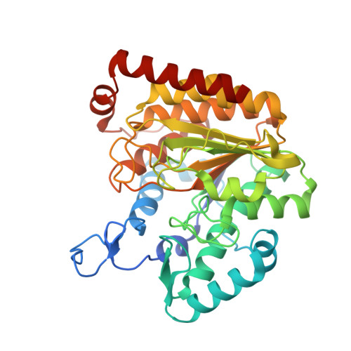
    Classical Zn 2+ -dependent deac(et)ylases play fundamental regulatory roles in life and are well characterized in eukaryotes regarding their structures, substrates and physiological roles. In bacteria, however, classical deacylases are less well understood. We construct a Generalized Profile (GP) and identify thousands of uncharacterized classical deacylases in bacteria, which are grouped into five clusters. Systematic structural and functional characterization of representative enzymes from each cluster reveal high functional diversity, including polyamine deacylases and protein deacylases with various acyl-chain type preferences. These data are supported by multiple crystal structures of enzymes from different clusters. Through this extensive analysis, we define the structural requirements of substrate selectivity, and discovered bacterial de-D-/L-lactylases and long-chain deacylases. Importantly, bacterial deacylases are inhibited by archetypal HDAC inhibitors, as supported by co-crystal structures with the inhibitors SAHA and TSA, and setting the ground for drug repurposing strategies to fight bacterial infections. Thus, we provide a systematic structure-function analysis of classical deacylases in bacteria and reveal the basis of substrate specificity, acyl-chain preference and inhibition.


  • Organizational Affiliation
    • Department Synthetic and Structural Biochemistry, Institute of Biochemistry, University of Greifswald, Greifswald, Germany.

Macromolecule Content 

  • Total Structure Weight: 334.75 kDa 
  • Atom Count: 25,009 
  • Modeled Residue Count: 2,960 
  • Deposited Residue Count: 3,040 
  • Unique protein chains: 1

Macromolecules

Find similar proteins by:|  3D Structure
Entity ID: 1
MoleculeChains  Sequence LengthOrganismDetailsImage
Propanil hydrolase
A, B, C, D, E
A, B, C, D, E, F, G, H
380Sphingomonas sp. Y57Mutation(s): 0 
Gene Names: prpH
UniProt
Find proteins for A0ACD6B8L2 (Sphingomonas sp. Y57)
Explore A0ACD6B8L2 
Go to UniProtKB:  A0ACD6B8L2
Entity Groups
Sequence Clusters30% Identity50% Identity70% Identity90% Identity95% Identity100% Identity
UniProt GroupA0ACD6B8L2
Sequence Annotations
Expand
Reference Sequence

Small Molecules

Ligands 4 Unique
IDChains Name / Formula / InChI Key2D Diagram3D Interactions
BGC

Query on BGC


Download:Ideal Coordinates CCD File
AA [auth E]
EA [auth F]
IA [auth G]
J [auth A]
MA [auth H]
AA [auth E],
EA [auth F],
IA [auth G],
J [auth A],
MA [auth H],
N [auth B],
R [auth C],
S [auth C],
Z [auth E]
beta-D-glucopyranose
C6 H12 O6
WQZGKKKJIJFFOK-VFUOTHLCSA-N
ZN
(Subject of Investigation/LOI)

Query on ZN


Download:Ideal Coordinates CCD File
BA [auth E]
FA [auth F]
JA [auth G]
K [auth A]
NA [auth H]
BA [auth E],
FA [auth F],
JA [auth G],
K [auth A],
NA [auth H],
O [auth B],
T [auth C],
W [auth D]
ZINC ION
Zn
PTFCDOFLOPIGGS-UHFFFAOYSA-N
ACT

Query on ACT


Download:Ideal Coordinates CCD File
DA [auth F]
HA [auth G]
I [auth A]
LA [auth H]
M [auth B]
DA [auth F],
HA [auth G],
I [auth A],
LA [auth H],
M [auth B],
Q [auth C],
V [auth D],
Y [auth E]
ACETATE ION
C2 H3 O2
QTBSBXVTEAMEQO-UHFFFAOYSA-M
K

Query on K


Download:Ideal Coordinates CCD File
CA [auth E]
GA [auth F]
KA [auth G]
L [auth A]
OA [auth H]
CA [auth E],
GA [auth F],
KA [auth G],
L [auth A],
OA [auth H],
P [auth B],
U [auth C],
X [auth D]
POTASSIUM ION
K
NPYPAHLBTDXSSS-UHFFFAOYSA-N

Experimental Data & Validation

Experimental Data

  • Method: X-RAY DIFFRACTION
  • Resolution: 1.48 Å
  • R-Value Free:  0.199 (Depositor), 0.202 (DCC) 
  • R-Value Work:  0.173 (Depositor), 0.176 (DCC) 
  • R-Value Observed: 0.174 (Depositor) 
Space Group: P 1
Unit Cell:
Length ( Å )Angle ( ˚ )
a = 103.861α = 113.603
b = 103.698β = 92.995
c = 104.499γ = 120.605
Software Package:
Software NamePurpose
REFMACrefinement
XDSdata reduction
XDSdata scaling
PHASERphasing

Structure Validation

View Full Validation Report


Entry History 

& Funding Information

Deposition Data


Funding OrganizationLocationGrant Number
German Research Foundation (DFG)GermanyLA2984/6-1
German Research Foundation (DFG)GermanyLA2984/8-1

Revision History  (Full details and data files)

  • Version 1.0: 2024-11-06
    Type: Initial release
  • Version 1.1: 2024-11-13
    Changes: Database references