9GFJ | pdb_00009gfj

Crystal structure of ASO binding Fab fragment with ASO143


Experimental Data Snapshot

  • Method: X-RAY DIFFRACTION
  • Resolution: 1.48 Å
  • R-Value Free: 
    0.206 (Depositor), 0.205 (DCC) 
  • R-Value Work: 
    0.183 (Depositor), 0.183 (DCC) 
  • R-Value Observed: 
    0.184 (Depositor) 

Starting Model: experimental
View more details

wwPDB Validation 3D Report Full Report

Validation slider image for 9GFJ

Ligand Structure Quality Assessment 


This is version 1.1 of the entry. See complete history

Literature

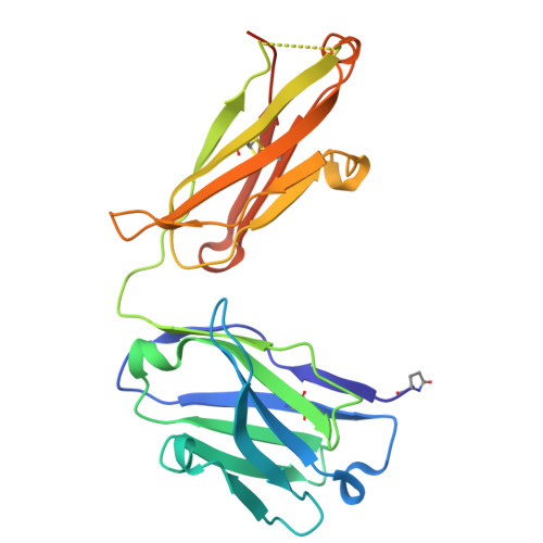
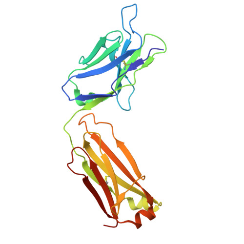
Improved targeted delivery of antisense oligonucleotide with an antibody mask.

Hsia, H.E.Zanini, C.Simonneau, C.Fraidling, J.Kraft, T.E.Mayer, K.Sommer, A.Indlekofer, A.Wirth, T.Benz, J.Georges, G.Langer, L.M.Gassner, C.Larraillet, V.Lohmann, S.Koller, E.Manso, M.Ravn, J.Hofer, K.Emrich, T.Niewohner, J.Schumacher, F.Brinkmann, U.

(2025) Nucleic Acids Res 53

  • DOI: https://doi.org/10.1093/nar/gkaf487
  • Primary Citation Related Structures: 
    9GF5, 9GFD, 9GFJ, 9GFL

  • PubMed Abstract: 

    Antisense oligonucleotides (ASOs) are synthetic nucleic acid strands designed to modulate gene expression by binding to RNA transcripts. In brain diseases, transferrin receptor (TfR)-targeting antibody-ASO conjugates have shown promise for brain delivery of ASOs via transcytosis in preclinical studies. This enables the more patient friendly intravenous or subcutaneous administration of the compounds. However, these conjugates can exhibit faster plasma clearance and different peripheral pharmacokinetic profiles due to ASO modifications, such as phosphorothioate (PS) linkages and locked nucleic acids (LNAs). In this study, we employed an antibody recognizing LNA- and PS-modified ASOs independent of the base sequences as a cloaking module to mitigate these issues. Using Kutzneria albida transglutaminase (KTG) technology and click chemistry, we generated TfR antibody-ASO conjugates with covalently or noncovalently incorporated ASO binders. Additionally, a noncovalent carrier antibody approach was explored. These conjugates and complexes with additional ASO binder(s) show improved TfR-targeted cellular uptake, undergo transcytosis in cellular blood-brain barrier models, show less nonspecific cellular accumulation, and anticipated ASO activity.


  • Organizational Affiliation
    • Roche Pharma Research and Early Development (pRED), Roche Innovation Center Munich, Roche Diagnostics GmbH, Penzberg82377, Germany.

Macromolecule Content 

  • Total Structure Weight: 49.69 kDa 
  • Atom Count: 3,865 
  • Modeled Residue Count: 428 
  • Deposited Residue Count: 445 
  • Unique protein chains: 2

Macromolecules

Find similar proteins by:|  3D Structure
Entity ID: 1
MoleculeChains  Sequence LengthOrganismDetailsImage
Fab fragment heavy chainA [auth H]226Homo sapiensMutation(s): 0 
Find similar proteins by:|  3D Structure
Entity ID: 2
MoleculeChains  Sequence LengthOrganismDetailsImage
Fab fragment light chainB [auth L]219Homo sapiensMutation(s): 0 

Small Molecules

Ligands 3 Unique
IDChains Name / Formula / InChI Key2D Diagram3D Interactions
A1IKV
(Subject of Investigation/LOI)

Query on A1IKV


Download:Ideal Coordinates CCD File
L
1-[(1~{S},3~{R},4~{R},7~{S})-7-[[(1~{R},3~{R},4~{R},7~{S})-7-[[(1~{R},3~{R},4~{R},7~{S})-3-(6-aminopurin-9-yl)-7-[[(1~{R},3~{R},4~{R},7~{S})-3-(4-azanyl-5-methyl-2-oxidanylidene-pyrimidin-1-yl)-7-oxidanyl-2,5-dioxabicyclo[2.2.1]heptan-1-yl]methoxy-sulfanyl-phosphoryl]oxy-2,5-dioxabicyclo[2.2.1]heptan-1-yl]methoxy-sulfanyl-phosphoryl]oxy-3-(4-azanyl-5-methyl-2-oxidanylidene-pyrimidin-1-yl)-2,5-dioxabicyclo[2.2.1]heptan-1-yl]methoxy-sulfanyl-phosphoryl]oxy-1-(hydroxymethyl)-2,5-dioxabicyclo[2.2.1]heptan-3-yl]-5-methyl-pyrimidine-2,4-dione
C44 H54 N13 O23 P3 S3
NZVZIQVDJJVXQQ-SSOWSIFGSA-N
EPE

Query on EPE


Download:Ideal Coordinates CCD File
I [auth L]4-(2-HYDROXYETHYL)-1-PIPERAZINE ETHANESULFONIC ACID
C8 H18 N2 O4 S
JKMHFZQWWAIEOD-UHFFFAOYSA-N
EDO

Query on EDO


Download:Ideal Coordinates CCD File
C [auth H]
D [auth H]
E [auth H]
F [auth H]
G [auth L]
C [auth H],
D [auth H],
E [auth H],
F [auth H],
G [auth L],
H [auth L],
J [auth L],
K [auth L]
1,2-ETHANEDIOL
C2 H6 O2
LYCAIKOWRPUZTN-UHFFFAOYSA-N
Modified Residues  1 Unique
IDChains TypeFormula2D DiagramParent
PCA
Query on PCA
A [auth H]L-PEPTIDE LINKINGC5 H7 N O3GLN

Experimental Data & Validation

Experimental Data

  • Method: X-RAY DIFFRACTION
  • Resolution: 1.48 Å
  • R-Value Free:  0.206 (Depositor), 0.205 (DCC) 
  • R-Value Work:  0.183 (Depositor), 0.183 (DCC) 
  • R-Value Observed: 0.184 (Depositor) 
Space Group: C 1 2 1
Unit Cell:
Length ( Å )Angle ( ˚ )
a = 127.539α = 90
b = 57.694β = 94.668
c = 63.046γ = 90
Software Package:
Software NamePurpose
PHENIXrefinement
XDSdata reduction
SADABSdata scaling
PHASERphasing

Structure Validation

View Full Validation Report


Ligand Structure Quality Assessment 


Entry History 

& Funding Information

Deposition Data


Funding OrganizationLocationGrant Number
Not funded--

Revision History  (Full details and data files)

  • Version 1.0: 2025-08-20
    Type: Initial release
  • Version 1.1: 2025-11-12
    Changes: Database references