9GCZ | pdb_00009gcz

XusB lipoprotein bound to ferric enterobactin


Experimental Data Snapshot

  • Method: X-RAY DIFFRACTION
  • Resolution: 1.50 Å
  • R-Value Free: 
    0.244 (Depositor), 0.244 (DCC) 
  • R-Value Work: 
    0.204 (Depositor), 0.204 (DCC) 
  • R-Value Observed: 
    0.206 (Depositor) 

Starting Model: experimental
View more details

wwPDB Validation 3D Report Full Report

Validation slider image for 9GCZ

Ligand Structure Quality Assessment 


This is version 1.1 of the entry. See complete history

Literature

Structural basis of iron piracy by human gut Bacteroides.

Silale, A.Soo, Y.L.Mark, H.Motz, R.N.Basle, A.Nolan, E.M.van den Berg, B.

(2025) bioRxiv 

  • DOI: https://doi.org/10.1101/2024.04.15.589501
  • Primary Citation Related Structures: 
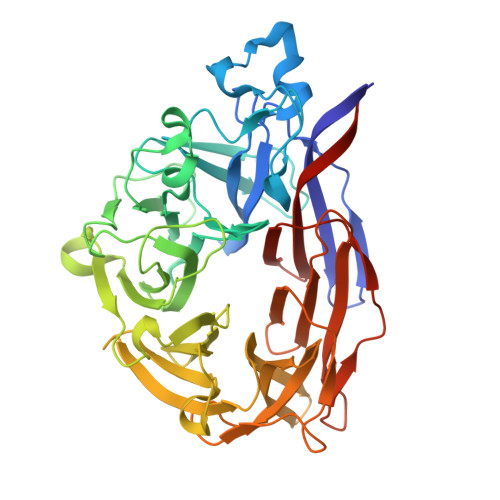
    9GAR, 9GBC, 9GCY, 9GCZ, 9HQ1, 9HQE, 9HQK

  • PubMed Abstract: 

    Iron is an essential element that can be growth-limiting in microbial communities, particularly those present within host organisms. To acquire iron, many bacteria secrete siderophores, secondary metabolites that chelate ferric iron. These iron chelates can be transported back into the cell via TonB-dependent transporters in the outer membrane, followed by intracellular liberation of the iron. Pathogenic Escherichia coli and Salmonella produce siderophores during gut infection. In response to iron starvation, the human gut symbiont Bacteroides thetaiotaomicron upregulates an iron piracy system, XusABC, which steals iron-bound siderophores from the invading pathogens. Here, we investigated the molecular details of xenosiderophore uptake across the outer membrane by the XusAB complex. Our crystal and cryogenic electron microscopy structures explain how the XusB lipoprotein recognises iron-bound xenosiderophores and passes them on to the XusA TonB-dependent transporter. Moreover, we show that Xus homologues can transport a variety of siderophores with different iron-chelating functional groups.


  • Organizational Affiliation
    • Biosciences Institute, Newcastle University, Framlington Place, Newcastle upon Tyne, NE2 4HH, United Kingdom.

Macromolecule Content 

  • Total Structure Weight: 98.65 kDa 
  • Atom Count: 7,583 
  • Modeled Residue Count: 850 
  • Deposited Residue Count: 880 
  • Unique protein chains: 1

Macromolecules

Find similar proteins by:|  3D Structure
Entity ID: 1
MoleculeChains  Sequence LengthOrganismDetailsImage
DUF4374 domain-containing protein
A, B
440Bacteroides thetaiotaomicron VPI-5482Mutation(s): 0 
Gene Names: BT_2064
UniProt
Find proteins for Q8A622 (Bacteroides thetaiotaomicron (strain ATCC 29148 / DSM 2079 / JCM 5827 / CCUG 10774 / NCTC 10582 / VPI-5482 / E50))
Explore Q8A622 
Go to UniProtKB:  Q8A622
Entity Groups
Sequence Clusters30% Identity50% Identity70% Identity90% Identity95% Identity100% Identity
UniProt GroupQ8A622
Sequence Annotations
Expand
Reference Sequence

Small Molecules

Ligands 6 Unique
IDChains Name / Formula / InChI Key2D Diagram3D Interactions
EB4
(Subject of Investigation/LOI)

Query on EB4


Download:Ideal Coordinates CCD File
E [auth A],
J [auth B]
N,N',N''-[(3S,7S,11S)-2,6,10-trioxo-1,5,9-trioxacyclododecane-3,7,11-triyl]tris(2,3-dihydroxybenzamide)
C30 H27 N3 O15
SERBHKJMVBATSJ-BZSNNMDCSA-N
CIT

Query on CIT


Download:Ideal Coordinates CCD File
C [auth A]CITRIC ACID
C6 H8 O7
KRKNYBCHXYNGOX-UHFFFAOYSA-N
SO4

Query on SO4


Download:Ideal Coordinates CCD File
H [auth B]SULFATE ION
O4 S
QAOWNCQODCNURD-UHFFFAOYSA-L
DMS

Query on DMS


Download:Ideal Coordinates CCD File
F [auth A]DIMETHYL SULFOXIDE
C2 H6 O S
IAZDPXIOMUYVGZ-UHFFFAOYSA-N
FE
(Subject of Investigation/LOI)

Query on FE


Download:Ideal Coordinates CCD File
D [auth A],
I [auth B]
FE (III) ION
Fe
VTLYFUHAOXGGBS-UHFFFAOYSA-N
NA

Query on NA


Download:Ideal Coordinates CCD File
G [auth A]SODIUM ION
Na
FKNQFGJONOIPTF-UHFFFAOYSA-N

Experimental Data & Validation

Experimental Data

  • Method: X-RAY DIFFRACTION
  • Resolution: 1.50 Å
  • R-Value Free:  0.244 (Depositor), 0.244 (DCC) 
  • R-Value Work:  0.204 (Depositor), 0.204 (DCC) 
  • R-Value Observed: 0.206 (Depositor) 
Space Group: P 1 21 1
Unit Cell:
Length ( Å )Angle ( ˚ )
a = 59.992α = 90
b = 48.18β = 96.787
c = 135.466γ = 90
Software Package:
Software NamePurpose
PHENIXrefinement
xia2data reduction
Aimlessdata scaling
PHENIXphasing

Structure Validation

View Full Validation Report


Ligand Structure Quality Assessment 


Entry History 

& Funding Information

Deposition Data


Funding OrganizationLocationGrant Number
Wellcome TrustUnited Kingdom214222/Z/18/Z

Revision History  (Full details and data files)

  • Version 1.0: 2025-08-13
    Type: Initial release
  • Version 1.1: 2025-09-17
    Changes: Database references