9G6W | pdb_00009g6w

L-SIGN CRD in complex with Man96.


Experimental Data Snapshot

  • Method: X-RAY DIFFRACTION
  • Resolution: 2.00 Å
  • R-Value Free: 
    0.290 (Depositor), 0.320 (DCC) 
  • R-Value Work: 
    0.228 (Depositor), 0.264 (DCC) 
  • R-Value Observed: 
    0.231 (Depositor) 

Starting Model: experimental
View more details

wwPDB Validation 3D Report Full Report

Validation slider image for 9G6W

Ligand Structure Quality Assessment 


This is version 1.1 of the entry. See complete history

Literature

Increasing the Chemical Space of L-SIGN Specific Glycomimetics.

Cavazzoli, G.Delaunay, C.Pollastri, S.Panzeri, A.Sattin, S.Thepaut, M.Belvisi, L.Fieschi, F.Bernardi, A.

(2025) J Med Chem 68: 22530-22546

  • DOI: https://doi.org/10.1021/acs.jmedchem.5c01448
  • Primary Citation Related Structures: 
    9G6W

  • PubMed Abstract: 

    Selective ligands for the C-type lectin receptor L-SIGN offer promising avenues in antiviral therapies and for tissue-specific delivery. We recently reported that a guanidine-bearing modified mannose glycomimetic, called Man84 , binds to L-SIGN with micromolar affinity and high-selectivity against the homologue lectin DC-SIGN. Here we describe a series of Man84 isosteres (ligands 2-11 ) that maintain or improve on this selectivity. The affinity of the ligands for L-SIGN, as well as their selectivity against DC-SIGN, were evaluated by Surface Plasmon Resonance inhibition assays using immobilized SARS-CoV-2 Spike protein. Compounds 4 , 5 and 9 were found to bind to L-SIGN with low micromolar affinity and 50-94-fold selectivity, thus matching or exceeding the performance of Man84 . The crystal structure of the L-SIGN CRD/ 4 complex was solved and highlighted the critical role of a bidentate H-bond interaction of the ligands with the side chain of E370 in L-SIGN.


  • Organizational Affiliation
    • Dipartimento di Chimica, Università degli Studi di Milano, via Golgi 19, Milano 20133, Italy.

Macromolecule Content 

  • Total Structure Weight: 32.83 kDa 
  • Atom Count: 2,304 
  • Modeled Residue Count: 256 
  • Deposited Residue Count: 274 
  • Unique protein chains: 1

Macromolecules

Find similar proteins by:|  3D Structure
Entity ID: 1
MoleculeChains  Sequence LengthOrganismDetailsImage
Isoform 1 of C-type lectin domain family 4 member M
A, B
137Homo sapiensMutation(s): 0 
Gene Names: CLEC4MCD209LCD209L1CD299
UniProt & NIH Common Fund Data Resources
Find proteins for Q9H2X3 (Homo sapiens)
Explore Q9H2X3 
Go to UniProtKB:  Q9H2X3
PHAROS:  Q9H2X3
GTEx:  ENSG00000104938 
Entity Groups
Sequence Clusters30% Identity50% Identity70% Identity90% Identity95% Identity100% Identity
UniProt GroupQ9H2X3
Sequence Annotations
Expand
Reference Sequence

Small Molecules

Ligands 4 Unique
IDChains Name / Formula / InChI Key2D Diagram3D Interactions
A1IIW
(Subject of Investigation/LOI)

Query on A1IIW


Download:Ideal Coordinates CCD File
C [auth A],
I [auth B]
(2~{R},3~{S},4~{R},5~{S},6~{S})-6-(2-chloroethyloxy)-2-(hydroxymethyl)-5-[4-[(imidazolidin-2-ylideneamino)methyl]-1,2,3-triazol-1-yl]oxane-3,4-diol
C14 H23 Cl N6 O5
SUTPIBBHKSRFDY-MLGHIDQZSA-N
TRS

Query on TRS


Download:Ideal Coordinates CCD File
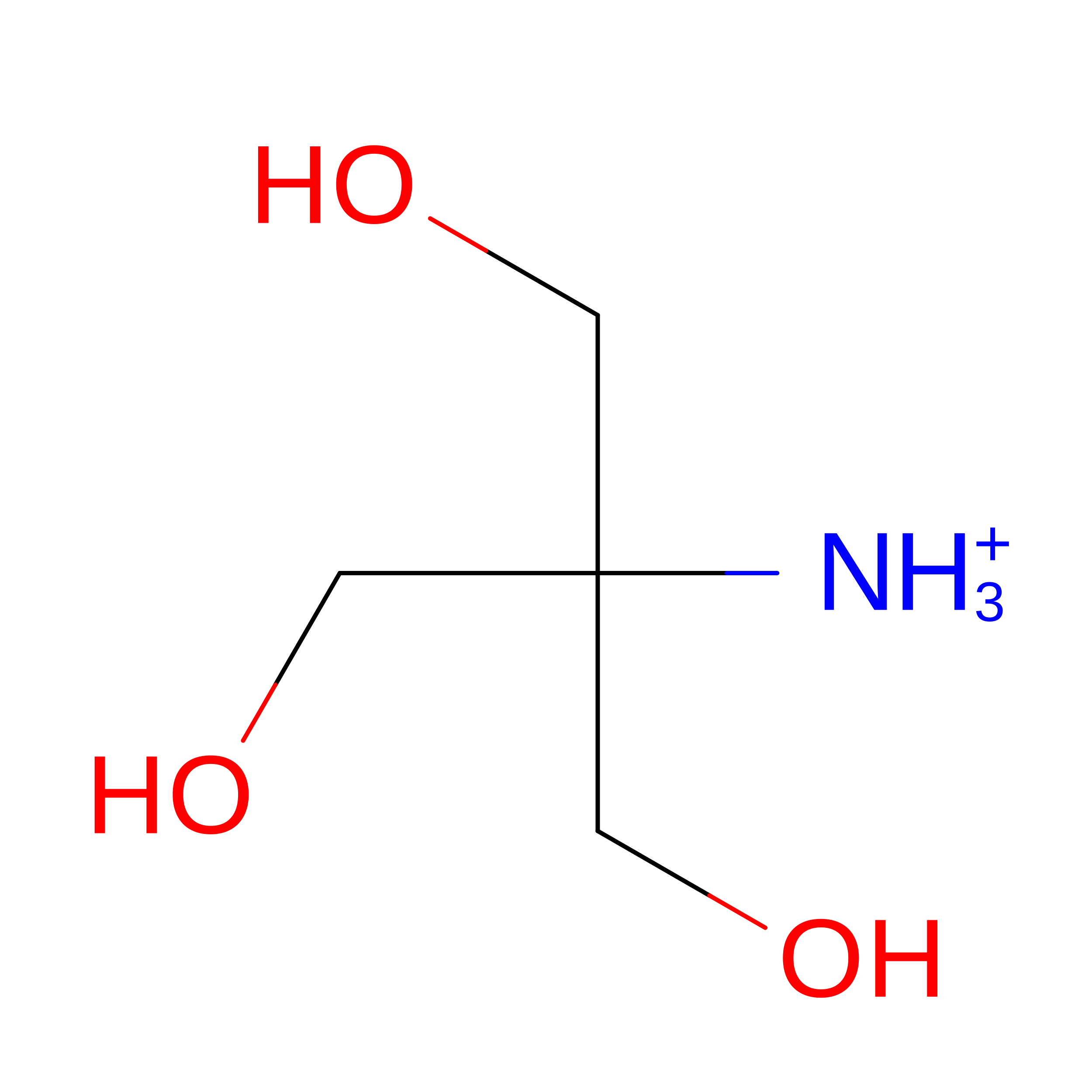
M [auth B]2-AMINO-2-HYDROXYMETHYL-PROPANE-1,3-DIOL
C4 H12 N O3
LENZDBCJOHFCAS-UHFFFAOYSA-O
CA

Query on CA


Download:Ideal Coordinates CCD File
D [auth A]
E [auth A]
F [auth A]
J [auth B]
K [auth B]
D [auth A],
E [auth A],
F [auth A],
J [auth B],
K [auth B],
L [auth B]
CALCIUM ION
Ca
BHPQYMZQTOCNFJ-UHFFFAOYSA-N
CL

Query on CL


Download:Ideal Coordinates CCD File
G [auth A],
H [auth A]
CHLORIDE ION
Cl
VEXZGXHMUGYJMC-UHFFFAOYSA-M

Experimental Data & Validation

Experimental Data

  • Method: X-RAY DIFFRACTION
  • Resolution: 2.00 Å
  • R-Value Free:  0.290 (Depositor), 0.320 (DCC) 
  • R-Value Work:  0.228 (Depositor), 0.264 (DCC) 
  • R-Value Observed: 0.231 (Depositor) 
Space Group: P 65
Unit Cell:
Length ( Å )Angle ( ˚ )
a = 105.72α = 90
b = 105.72β = 90
c = 59.11γ = 120
Software Package:
Software NamePurpose
XDSdata reduction
XSCALEdata scaling
MOLREPphasing
Cootmodel building
REFMACrefinement

Structure Validation

View Full Validation Report


Ligand Structure Quality Assessment 


Entry History 

& Funding Information

Deposition Data


Funding OrganizationLocationGrant Number
Grenoble Alliance for Integrated Structural Cell Biology (GRAL)FranceCBH-EUR-GS (ANR-17- EURE0003)
Ministero dell Universita e della RicercaItalyPRIN 20224LLK82

Revision History  (Full details and data files)

  • Version 1.0: 2025-07-30
    Type: Initial release
  • Version 1.1: 2026-09-16
    Changes: Database references