9G18 | pdb_00009g18

Structure of PslG with an iminosugar inhibitor


Experimental Data Snapshot

  • Method: X-RAY DIFFRACTION
  • Resolution: 1.50 Å
  • R-Value Free: 
    0.160 (Depositor), 0.160 (DCC) 
  • R-Value Work: 
    0.131 (Depositor), 0.131 (DCC) 

Starting Model: experimental
View more details

wwPDB Validation 3D Report Full Report

Validation slider image for 9G18

This is version 1.1 of the entry. See complete history

Literature

Bespoke Activity-Based Probes Reveal that the Pseudomonas aeruginosa Endoglycosidase, PslG, Is an Endo-beta-glucanase.

Ruijgrok, G.Offen, W.A.Pickles, I.B.Raju, D.Patsos, T.de Boer, C.Ofman, T.Rompa, J.van Oord, D.Dodson, E.J.Beekers, A.Voskuilen, T.Ferrari, M.Wu, L.Janssen, A.P.A.Codee, J.D.C.Howell, P.L.Davies, G.J.Overkleeft, H.S.

(2025) J Am Chem Soc 147: 8578-8586

  • DOI: https://doi.org/10.1021/jacs.4c16806
  • Primary Citation Related Structures: 
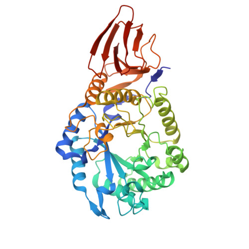
    9G17, 9G18

  • PubMed Abstract: 

    During infection, the human opportunistic pathogen Pseudomonas aeruginosa forms protective biofilms, whose matrix consists of proteins, nucleic acids, and polysaccharides such as alginate, Psl, and Pel. Psl, a polymeric pentasaccharide composed of mannose, rhamnose, and glucose, is produced during the early stages of biofilm formation, serving as a protective barrier against antibiotics and the immune system. The Psl biosynthesis gene cluster, besides encoding various glycosyltransferases, also includes an endoglycosidase, PslG. Here, we show, by activity-based protein profiling, structural studies on enzyme-inhibitor complexes, and defined substrate processing, that PslG is not, as previously suggested, an endo-β-mannosidase but instead a retaining endo-β-glucosidase. This insight allows the design of both competitive and covalent PslG inhibitors, as we show for repeating pentasaccharide mimetics featuring either a reducing end deoxynojirimycin or cyclophellitol moiety. This work provides valuable tools to deepen the understanding of Psl biosynthesis, its function in biofilm formation, and its contribution to antibiotic resistance. We demonstrate the enzyme's actual endo-β-glucosidase activity, a means to monitor PslG activity in P. aeruginosa biofilms, and a blueprint for inhibitor design.


  • Organizational Affiliation
    • Leiden Institute of Chemistry, Leiden University, 2300 RA Leiden, The Netherlands.

Macromolecule Content 

  • Total Structure Weight: 48.74 kDa 
  • Atom Count: 4,137 
  • Modeled Residue Count: 412 
  • Deposited Residue Count: 416 
  • Unique protein chains: 1

Macromolecules

Find similar proteins by:|  3D Structure
Entity ID: 1
MoleculeChains  Sequence LengthOrganismDetailsImage
PslG416Pseudomonas aeruginosaMutation(s): 0 
Gene Names: pslGPA2237
UniProt
Find proteins for Q9I1N2 (Pseudomonas aeruginosa (strain ATCC 15692 / DSM 22644 / CIP 104116 / JCM 14847 / LMG 12228 / 1C / PRS 101 / PAO1))
Explore Q9I1N2 
Go to UniProtKB:  Q9I1N2
Entity Groups
Sequence Clusters30% Identity50% Identity70% Identity90% Identity95% Identity100% Identity
UniProt GroupQ9I1N2
Sequence Annotations
Expand
Reference Sequence

Oligosaccharides

Help  
Entity ID: 2
MoleculeChains Length2D Diagram GlycosylationD Interactions
alpha-D-mannopyranose-(1-2)-beta-D-mannopyranose-(1-3)-beta-D-mannopyranose-(1-3)-alpha-L-rhamnopyranose-(1-3)-1-DEOXYNOJIRIMYCIN
B
5N/A

Small Molecules

Ligands 2 Unique
IDChains Name / Formula / InChI Key2D Diagram3D Interactions
GOL

Query on GOL


Download:Ideal Coordinates CCD File
C [auth A],
D [auth A],
E [auth A],
F [auth A],
G [auth A]
GLYCEROL
C3 H8 O3
PEDCQBHIVMGVHV-UHFFFAOYSA-N
CL

Query on CL


Download:Ideal Coordinates CCD File
H [auth A],
I [auth A],
J [auth A]
CHLORIDE ION
Cl
VEXZGXHMUGYJMC-UHFFFAOYSA-M

Experimental Data & Validation

Experimental Data

  • Method: X-RAY DIFFRACTION
  • Resolution: 1.50 Å
  • R-Value Free:  0.160 (Depositor), 0.160 (DCC) 
  • R-Value Work:  0.131 (Depositor), 0.131 (DCC) 
Space Group: P 65
Unit Cell:
Length ( Å )Angle ( ˚ )
a = 97.604α = 90
b = 97.604β = 90
c = 119.852γ = 120
Software Package:
Software NamePurpose
REFMACrefinement
DIALSdata reduction
Aimlessdata scaling
PHASERphasing

Structure Validation

View Full Validation Report


Entry History 

& Funding Information

Deposition Data


Funding OrganizationLocationGrant Number
European Research Council (ERC)European Union951231

Revision History  (Full details and data files)

  • Version 1.0: 2025-03-05
    Type: Initial release
  • Version 1.1: 2025-03-26
    Changes: Database references