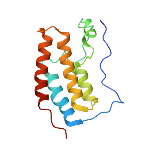
9FXP

Crystal structure of BRD4 BD1 with DI00626383.


Experimental Data Snapshot

  • Method: X-RAY DIFFRACTION
  • Resolution: 1.88 Å
  • R-Value Free: 0.218 
  • R-Value Work: 0.181 
  • R-Value Observed: 0.182 

Starting Model: experimental
View more details

wwPDB Validation   3D Report Full Report


Ligand Structure Quality Assessment 


This is version 1.1 of the entry. See complete history


Literature

Probing Protein-Ligand Methyl-pi Interaction Geometries through Chemical Shift Measurements of Selectively Labeled Methyl Groups.

Beier, A.Platzer, G.Hofurthner, T.Ptaszek, A.L.Lichtenecker, R.J.Geist, L.Fuchs, J.E.McConnell, D.B.Mayer, M.Konrat, R.

(2024) J Med Chem 67: 13187-13196

  • DOI: https://doi.org/10.1021/acs.jmedchem.4c01128
  • Primary Citation of Related Structures:  
    9FWX, 9FXP

  • PubMed Abstract: 

    Fragment-based drug design is heavily dependent on the optimization of initial low-affinity binders. Herein we introduce an approach that uses selective labeling of methyl groups in leucine and isoleucine side chains to directly probe methyl-π contacts, one of the most prominent forms of interaction between proteins and small molecules. Using simple NMR chemical shift perturbation experiments with selected BRD4-BD1 binders, we find good agreement with a commonly used model of the ring-current effect as well as the overall interaction geometries extracted from the Protein Data Bank. By combining both interaction geometries and chemical shift calculations as fit quality criteria, we can position dummy aromatic rings into an AlphaFold model of the protein of interest. The proposed method can therefore provide medicinal chemists with important information about binding geometries of small molecules in fast and iterative matter, even in the absence of high-resolution experimental structures.


  • Organizational Affiliation

    Christian Doppler Laboratory for High-Content Structural Biology and Biotechnology, Department of Structural and Computational Biology, Max Perutz Laboratories, University of Vienna, Campus Vienna Biocenter 5, 1030 Vienna, Austria.


Macromolecules
Find similar proteins by:  (by identity cutoff)  |  3D Structure
Entity ID: 1
MoleculeChains Sequence LengthOrganismDetailsImage
Bromodomain-containing protein 4127Homo sapiensMutation(s): 0 
Gene Names: BRD4HUNK1
UniProt & NIH Common Fund Data Resources
Find proteins for O60885 (Homo sapiens)
Explore O60885 
Go to UniProtKB:  O60885
PHAROS:  O60885
GTEx:  ENSG00000141867 
Entity Groups  
Sequence Clusters30% Identity50% Identity70% Identity90% Identity95% Identity100% Identity
UniProt GroupO60885
Sequence Annotations
Expand
  • Reference Sequence
Small Molecules
Ligands 1 Unique
IDChains Name / Formula / InChI Key2D Diagram3D Interactions
5WX (Subject of Investigation/LOI)
Query on 5WX

Download Ideal Coordinates CCD File 
B [auth A]4-methoxy-1,2-benzoxazol-3-amine
C8 H8 N2 O2
YFNIGSJHKGKXEQ-UHFFFAOYSA-N
Experimental Data & Validation

Experimental Data

  • Method: X-RAY DIFFRACTION
  • Resolution: 1.88 Å
  • R-Value Free: 0.218 
  • R-Value Work: 0.181 
  • R-Value Observed: 0.182 
  • Space Group: P 21 21 21
Unit Cell:
Length ( Å )Angle ( ˚ )
a = 36.99α = 90
b = 43.98β = 90
c = 78.23γ = 90
Software Package:
Software NamePurpose
BUSTERrefinement
SCALAdata scaling
XDSdata reduction
BUSTERphasing

Structure Validation

View Full Validation Report


Ligand Structure Quality Assessment 


Entry History & Funding Information

Deposition Data


Funding OrganizationLocationGrant Number
Not funded--

Revision History  (Full details and data files)

  • Version 1.0: 2024-08-07
    Type: Initial release
  • Version 1.1: 2024-08-21
    Changes: Database references