9FTB | pdb_00009ftb

Aeromonas caviae CMP-Pse5A7Ac Synthetase


Experimental Data Snapshot

  • Method: X-RAY DIFFRACTION
  • Resolution: 1.90 Å
  • R-Value Free: 
    0.241 (Depositor), 0.236 (DCC) 
  • R-Value Work: 
    0.192 (Depositor), 0.190 (DCC) 

Starting Model: in silico
View more details

wwPDB Validation 3D Report Full Report

Validation slider image for 9FTB

This is version 1.2 of the entry. See complete history

Literature

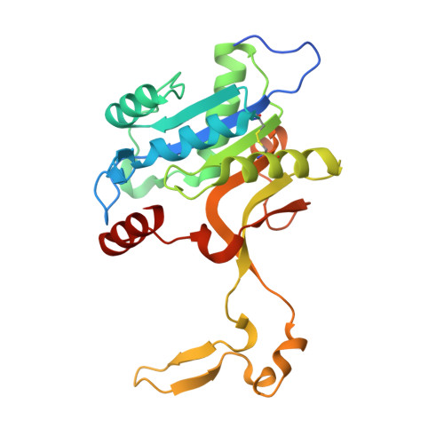
Structural dissection of the CMP-pseudaminic acid synthetase, PseF.

Keenan, T.Cowan, A.R.Flack, E.K.P.Hatton, N.E.Walklett, A.J.Thomas, G.H.Hemsworth, G.R.Fascione, M.A.

(2024) Structure 32: 2399

  • DOI: https://doi.org/10.1016/j.str.2024.09.017
  • Primary Citation Related Structures: 
    9FTB, 9FTC

  • PubMed Abstract: 

    Pseudaminic acid is a non-mammalian sugar found in the surface glycoconjugates of many bacteria, including several human pathogens, and is a virulence factor thought to facilitate immune evasion. The final step in the biosynthesis of the nucleotide activated form of the sugar, CMP-Pse5Ac7Ac is performed by a CMP-Pse5Ac7Ac synthetase (PseF). Here we present the biochemical and structural characterization of PseF from Aeromonas caviae (AcPseF), with AcPseF displaying metal-dependent activity over a broad pH and temperature range. Upon binding to CMP-Pse5Ac7Ac, AcPseF undergoes dynamic movements akin to other CMP-ulosonic acid synthetases. The enzyme clearly discriminates Pse5Ac7Ac from other ulosonic acids, through active site interactions with side-chain functional groups and by positioning the molecule in a hydrophobic pocket. Finally, we show that AcPseF binds the CMP-Pse5Ac7Ac side chain in the lowest energy conformation, a trend that we observed in the structures of other enzymes of this class.


  • Organizational Affiliation
    • Department of Chemistry, University of York, York YO10 5DD, UK.

Macromolecule Content 

  • Total Structure Weight: 28.01 kDa 
  • Atom Count: 1,990 
  • Modeled Residue Count: 234 
  • Deposited Residue Count: 248 
  • Unique protein chains: 1

Macromolecules

Find similar proteins by:|  3D Structure
Entity ID: 1
MoleculeChains  Sequence LengthOrganismDetailsImage
NeuA248Aeromonas caviaeMutation(s): 0 
Gene Names: neuA
UniProt
Find proteins for Q9R9S4 (Aeromonas caviae)
Explore Q9R9S4 
Go to UniProtKB:  Q9R9S4
Entity Groups
Sequence Clusters30% Identity50% Identity70% Identity90% Identity95% Identity100% Identity
UniProt GroupQ9R9S4
Sequence Annotations
Expand
Reference Sequence

Experimental Data & Validation

Experimental Data

  • Method: X-RAY DIFFRACTION
  • Resolution: 1.90 Å
  • R-Value Free:  0.241 (Depositor), 0.236 (DCC) 
  • R-Value Work:  0.192 (Depositor), 0.190 (DCC) 
Space Group: P 65 2 2
Unit Cell:
Length ( Å )Angle ( ˚ )
a = 109.527α = 90
b = 109.527β = 90
c = 87.063γ = 120
Software Package:
Software NamePurpose
REFMACrefinement
Aimlessdata scaling
PHASERphasing
XDSdata reduction

Structure Validation

View Full Validation Report


Entry History 

& Funding Information

Deposition Data


Funding OrganizationLocationGrant Number
European Research Council (ERC)European Union101044024

Revision History  (Full details and data files)

  • Version 1.0: 2024-10-09
    Type: Initial release
  • Version 1.1: 2024-10-30
    Changes: Database references
  • Version 1.2: 2024-12-18
    Changes: Database references