9FB8 | pdb_00009fb8

XChem refined PIF1-x0076 complex


Experimental Data Snapshot

  • Method: X-RAY DIFFRACTION
  • Resolution: 1.73 Å
  • R-Value Free: 
    0.211 (Depositor), 0.192 (DCC) 
  • R-Value Work: 
    0.173 (Depositor) 

Starting Model: experimental
View more details

wwPDB Validation 3D Report Full Report

Validation slider image for 9FB8

Ligand Structure Quality Assessment 


This is version 1.2 of the entry. See complete history

Literature

Structure-based discovery of first inhibitors targeting the helicase activity of human PIF1.

Wever, M.J.A.Scommegna, F.R.Egea-Rodriguez, S.Dehghani-Tafti, S.Brandao-Neto, J.Poisson, J.F.Helfrich, I.Antson, A.A.Rodeschini, V.Bax, B.Roche, D.Sanders, C.M.

(2024) Nucleic Acids Res 52: 12616-12632

  • DOI: https://doi.org/10.1093/nar/gkae897
  • Primary Citation Related Structures: 
    9FB8, 9FI9

  • PubMed Abstract: 

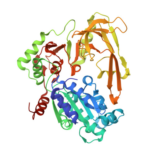
    PIF1 is a conserved helicase and G4 DNA binding and unwinding enzyme, with roles in genome stability. Human PIF1 (hPIF1) is poorly understood, but its functions can become critical for tumour cell survival during oncogene-driven replication stress. Here we report the discovery, via an X-ray crystallographic fragment screen (XChem), of hPIF1 DNA binding and unwinding inhibitors. A structure was obtained with a 4-phenylthiazol-2-amine fragment bound in a pocket between helicase domains 2A and 2B, with additional contacts to Valine 258 from domain 1A. The compound makes specific interactions, notably through Leucine 548 and Alanine 551, that constrain conformational adjustments between domains 2A and 2B, previously linked to ATP hydrolysis and DNA unwinding. We next synthesized a range of related compounds and characterized their effects on hPIF1 DNA-binding and helicase activity in vitro, expanding the structure activity relationship (SAR) around the initial hit. A systematic analysis of clinical cancer databases is also presented here, supporting the notion that hPIF1 upregulation may represent a specific cancer cell vulnerability. The research demonstrates that hPIF1 is a tractable target through 4-phenylthiazol-2-amine derivatives as inhibitors of its helicase action, setting a foundation for creation of a novel class of anti-cancer therapeutics.


  • Organizational Affiliation
    • Edelris, Bioparc, Bioserra 1 Building, 69008 Lyon, France.

Macromolecule Content 

  • Total Structure Weight: 46.65 kDa 
  • Atom Count: 3,675 
  • Modeled Residue Count: 418 
  • Deposited Residue Count: 419 
  • Unique protein chains: 1

Macromolecules

Find similar proteins by:|  3D Structure
Entity ID: 1
MoleculeChains  Sequence LengthOrganismDetailsImage
ATP-dependent DNA helicase PIF1419Homo sapiensMutation(s): 0 
Gene Names: PIF1C15orf20
EC: 3.6.4.12 (PDB Primary Data), 5.6.2.3 (UniProt)
UniProt & NIH Common Fund Data Resources
Find proteins for Q9H611 (Homo sapiens)
Explore Q9H611 
Go to UniProtKB:  Q9H611
PHAROS:  Q9H611
GTEx:  ENSG00000140451 
Entity Groups
Sequence Clusters30% Identity50% Identity70% Identity90% Identity95% Identity100% Identity
UniProt GroupQ9H611
Sequence Annotations
Expand
Reference Sequence

Small Molecules

Ligands 4 Unique
IDChains Name / Formula / InChI Key2D Diagram3D Interactions
ANP

Query on ANP


Download:Ideal Coordinates CCD File
B [auth A]PHOSPHOAMINOPHOSPHONIC ACID-ADENYLATE ESTER
C10 H17 N6 O12 P3
PVKSNHVPLWYQGJ-KQYNXXCUSA-N
O0J
(Subject of Investigation/LOI)

Query on O0J


Download:Ideal Coordinates CCD File
D [auth A],
E [auth A]
N-[4-(2-amino-1,3-thiazol-4-yl)phenyl]acetamide
C11 H11 N3 O S
VBBNSESFUHRMJU-UHFFFAOYSA-N
DMS

Query on DMS


Download:Ideal Coordinates CCD File
F [auth A]DIMETHYL SULFOXIDE
C2 H6 O S
IAZDPXIOMUYVGZ-UHFFFAOYSA-N
MG

Query on MG


Download:Ideal Coordinates CCD File
C [auth A]MAGNESIUM ION
Mg
JLVVSXFLKOJNIY-UHFFFAOYSA-N

Experimental Data & Validation

Experimental Data

  • Method: X-RAY DIFFRACTION
  • Resolution: 1.73 Å
  • R-Value Free:  0.211 (Depositor), 0.192 (DCC) 
  • R-Value Work:  0.173 (Depositor) 
Space Group: C 2 2 21
Unit Cell:
Length ( Å )Angle ( ˚ )
a = 73.6α = 90
b = 143.714β = 90
c = 77.238γ = 90
Software Package:
Software NamePurpose
REFMACrefinement
DIALSdata reduction
DIALSdata scaling
REFMACphasing

Structure Validation

View Full Validation Report


Ligand Structure Quality Assessment 


Entry History 

& Funding Information

Deposition Data


Funding OrganizationLocationGrant Number
Diamond Light SourceUnited KingdomLB19204

Revision History  (Full details and data files)

  • Version 1.0: 2024-09-18
    Type: Initial release
  • Version 1.1: 2024-10-30
    Changes: Database references, Structure summary
  • Version 1.2: 2024-11-20
    Changes: Database references