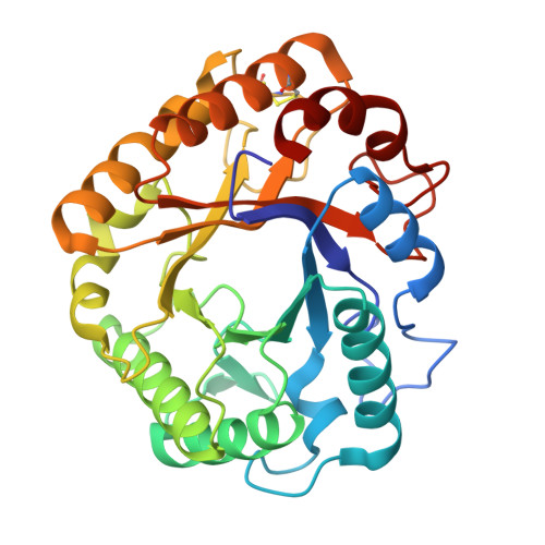
9F0N | pdb_00009f0n

Crystal structure of Ta_Cel5A E133A variant, apoform


Experimental Data Snapshot

  • Method: X-RAY DIFFRACTION
  • Resolution: 1.18 Å
  • R-Value Free: 
    0.159 (Depositor), 0.159 (DCC) 
  • R-Value Work: 
    0.149 (Depositor), 0.149 (DCC) 
  • R-Value Observed: 
    0.149 (Depositor) 

Starting Model: experimental
View more details

wwPDB Validation 3D Report Full Report

Validation slider image for 9F0N

This is version 1.1 of the entry. See complete history

Literature

Definition of the catalytic cleft and product profile of a GH5_5 cellulase from Thermoascus aurantiacus.

Collet, L.Denys, A.Oudjama, Y.Sifennasr, K.Vander Wauven, C.Dutoit, R.

(2026) Acta Crystallogr D Struct Biol 

  • DOI: https://doi.org/10.1107/S2059798326003578
  • Primary Citation Related Structures: 
    7R28, 7R29, 7R2A, 7R2C, 7R2D, 9F0N

  • PubMed Abstract: 

    Glycoside hydrolases (GHs) achieve glycan breakdown through glycosidic bond hydrolysis. Some retaining GHs can also transglycosylate, which could be useful for glycosynthesis. Improving GHs for either glycan degradation or synthesis requires a deep understanding of the residues involved in catalysis and substrate binding. This study characterizes in detail the activity and structure of Ta_Cel5A, a cellulase in glycoside hydrolase family 5, subfamily 5 (GH5_5) from the thermophilic ascomycete Thermoascus aurantiacus. While its hydrolytic activity was confirmed, Ta_Cel5A was also found to exhibit a weak transglycosylase activity with cellopentaose as a substrate. Transglycosylation products were detected within the first minutes of reaction at 25°C, far below its optimal temperature. The structures of catalytically impaired variants were solved in complex with oligosaccharides. The entire catalytic cleft was defined, consisting of seven glucose-binding subsites, five negative subsites and two positive subsites, from the nonreducing end to the reducing end. The fifth negative subsite could not be inferred in silico, showing the limitation in predicting distal subsites based on structural analogy. The structure of the glycosyl-enzyme intermediate was also obtained, revealing the displacement of key residues in the active site. The covalent binding of a glycosidic molecule triggers a major displacement of the nucleophilic residue, Glu244, changing its interaction network. The acid/base residue, Glu133, and a conserved tyrosine residue, Tyr201, are also displaced during glycosyl-enzyme intermediate formation, hinting at their role in the deglycosylation step.


  • Organizational Affiliation
    • LABIRIS, 1 Avenue Emile Gryzon, 1070 Brussels, Belgium.

Macromolecule Content 

  • Total Structure Weight: 68.63 kDa 
  • Atom Count: 5,879 
  • Modeled Residue Count: 609 
  • Deposited Residue Count: 612 
  • Unique protein chains: 1

Macromolecules

Find similar proteins by:|  3D Structure
Entity ID: 1
MoleculeChains  Sequence LengthOrganismDetailsImage
cellulase
A, B
306Thermoascus aurantiacus ATCC 26904Mutation(s): 1 
Gene Names: eg1
EC: 3.2.1.4
UniProt
Find proteins for Q8TG26 (Thermoascus aurantiacus)
Explore Q8TG26 
Go to UniProtKB:  Q8TG26
Entity Groups
Sequence Clusters30% Identity50% Identity70% Identity90% Identity95% Identity100% Identity
UniProt GroupQ8TG26
Sequence Annotations
Expand
Reference Sequence

Small Molecules

Ligands 2 Unique
IDChains Name / Formula / InChI Key2D Diagram3D Interactions
SO4

Query on SO4


Download:Ideal Coordinates CCD File
F [auth B],
G [auth B],
H [auth B]
SULFATE ION
O4 S
QAOWNCQODCNURD-UHFFFAOYSA-L
GOL

Query on GOL


Download:Ideal Coordinates CCD File
C [auth A]
D [auth A]
E [auth A]
I [auth B]
J [auth B]
C [auth A],
D [auth A],
E [auth A],
I [auth B],
J [auth B],
K [auth B],
L [auth B],
M [auth B]
GLYCEROL
C3 H8 O3
PEDCQBHIVMGVHV-UHFFFAOYSA-N

Experimental Data & Validation

Experimental Data

  • Method: X-RAY DIFFRACTION
  • Resolution: 1.18 Å
  • R-Value Free:  0.159 (Depositor), 0.159 (DCC) 
  • R-Value Work:  0.149 (Depositor), 0.149 (DCC) 
  • R-Value Observed: 0.149 (Depositor) 
Space Group: P 21 21 21
Unit Cell:
Length ( Å )Angle ( ˚ )
a = 76.56α = 90
b = 84.92β = 90
c = 89.27γ = 90
Software Package:
Software NamePurpose
PHENIXrefinement
XSCALEdata scaling
XDSdata reduction
PHASERphasing

Structure Validation

View Full Validation Report


Entry History 

& Funding Information

Deposition Data

  • Released Date: 2025-04-30 
  • Deposition Author(s): Dutoit, R.

Funding OrganizationLocationGrant Number
Other governmentBAG 20210875

Revision History  (Full details and data files)

  • Version 1.0: 2025-04-30
    Type: Initial release
  • Version 1.1: 2026-05-27
    Changes: Database references