A split-site E3 ligase mechanism enables ZNFX1 to ubiquitinate and cluster single-stranded RNA into ubiquitin-coated nucleoprotein particles.
Grabarczyk, D.B., Aird, E.J., Reznikow, V., Kirchgatterer, P.C., Ehrmann, J.F., Kurzbauer, R., Bell, L.E., Kellner, M.J., Aggarwal, R., Schleiffer, A., Faas, V., Deszcz, L., Meinhart, A., Versteeg, G.A., Penninger, J.M., Stelzl, L.S., Gaidt, M.M., Tessmer, I., Corn, J.E., Clausen, T.(2025) Cell 188: 5995
- PubMed: 40876457 Search on PubMed
- DOI: https://doi.org/10.1016/j.cell.2025.08.006
- Primary Citation Related Structures:
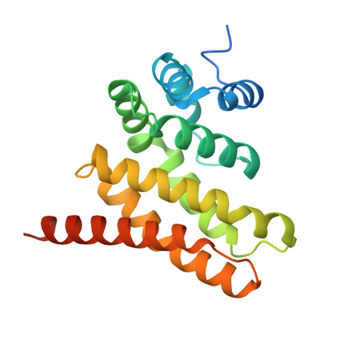
9EVE, 9Q9Z - PubMed Abstract:
Eukaryotic cells use a multi-layered immune response to combat intracellular pathogens. The ubiquitin ligase ZNFX1 has emerged as a crucial yet little understood player that regulates the immune response while protecting against RNA viruses. Our study unveils the molecular mechanism of ZNFX1, mediated by the joint activity of a helicase serving as a nucleic acid sensor and a non-conventional E3 module featuring a split active site. We demonstrate that single-stranded RNA stimulates E3 activity by fostering dimerization of ZNFX1 subunits that translocate along nucleic acid tracks. Juxtaposed E3 domains complement each other, leading to the ubiquitination of ZNFX1 itself and engaged RNA molecules, while clustering nucleic acids into dense nucleoprotein particles. We show that the E3 ligase activity of ZNFX1 protects cells during an immune response and propose that ubiquitin-coated particles formed by ZNFX1 represent part of an ancient mechanism to regulate both foreign and host RNA in the cell.
- Research Institute of Molecular Pathology, Vienna BioCenter, 1030 Vienna, Austria. Electronic address: daniel.grabarczyk@imp.ac.at.
Organizational Affiliation: