9EO2 | pdb_00009eo2

X-ray structure of the adduct formed upon reaction of picoplatin with lysozyme (structure B)


Experimental Data Snapshot

  • Method: X-RAY DIFFRACTION
  • Resolution: 1.36 Å
  • R-Value Free: 
    0.208 (Depositor), 0.214 (DCC) 
  • R-Value Work: 
    0.173 (Depositor), 0.182 (DCC) 

Starting Model: experimental
View more details

wwPDB Validation 3D Report Full Report

Validation slider image for 9EO2

This is version 1.2 of the entry. See complete history

Literature

Picoplatin binding to proteins: X-ray structures and mass spectrometry data on the adducts with lysozyme and ribonuclease A.

Ferraro, G.Lyckova, T.Massai, L.Starha, P.Messori, L.Merlino, A.

(2024) Dalton Trans 53: 8535-8540

  • DOI: https://doi.org/10.1039/d4dt00773e
  • Primary Citation Related Structures: 
    9ENZ, 9EO2, 9EO5, 9EO8

  • PubMed Abstract: 

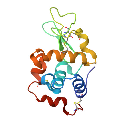
    The reactivity of the anticancer drug picoplatin ( cis -amminedichlorido(2-methylpyridine)platinum(II) complex) with the model proteins hen egg white lysozyme (HEWL) and bovine pancreatic ribonuclease (RNase A) was investigated by electrospray ionisation mass spectrometry (ESI MS) and X-ray crystallography. The data were compared with those previously obtained for the adducts of these proteins with cisplatin, carboplatin and oxaliplatin under the same experimental conditions. ESI-MS data show binding of Pt to both proteins, with fragments retaining the 2-methylpyridine ligand and, possibly, a chloride ion. X-ray crystallography identifies different binding sites on the two proteins, highlighting a different behaviour of picoplatin in the absence or presence of dimethyl sulfoxide (DMSO). Metal-containing fragments bind to HEWL close to the side chains of His15, Asp18, Asp119 and both Lys1 and Glu7, whereas they bind to RNase A on the side chain of His12, Met29, His48, Asp53, Met79, His105 and His119. The data suggest that the presence of DMSO favours the loss of 2-methylpyridine and alters the ability of the Pt compound to bind to the two proteins. With both proteins, picoplatin appears to behave similarly to cisplatin and carboplatin when dissolved in DMSO, whereas it behaves more like oxaliplatin in the absence of the coordinating solvent. This study provides important insights into the pharmacological profile of picoplatin and supports the conclusion that coordinating solvents should not be used to evaluate the biological activities of Pt-based drugs.


  • Organizational Affiliation
    • Department of Chemical Sciences, University of Naples Federico II, Complesso universitario di Monte Sant'Angelo, via Cinthia, 21, 80126, Naples, Italy. antonello.merlino@unina.it.

Macromolecule Content 

  • Total Structure Weight: 15.24 kDa 
  • Atom Count: 1,230 
  • Modeled Residue Count: 129 
  • Deposited Residue Count: 129 
  • Unique protein chains: 1

Macromolecules

Find similar proteins by:|  3D Structure
Entity ID: 1
MoleculeChains  Sequence LengthOrganismDetailsImage
Lysozyme CA [auth AAA]129Gallus gallusMutation(s): 0 
EC: 3.2.1.17
UniProt
Find proteins for P00698 (Gallus gallus)
Explore P00698 
Go to UniProtKB:  P00698
Entity Groups
Sequence Clusters30% Identity50% Identity70% Identity90% Identity95% Identity100% Identity
UniProt GroupP00698
Sequence Annotations
Expand
Reference Sequence

Small Molecules

Ligands 4 Unique
IDChains Name / Formula / InChI Key2D Diagram3D Interactions
PT
(Subject of Investigation/LOI)

Query on PT


Download:Ideal Coordinates CCD File
J [auth AAA],
K [auth AAA]
PLATINUM (II) ION
Pt
HRGDZIGMBDGFTC-UHFFFAOYSA-N
GOL

Query on GOL


Download:Ideal Coordinates CCD File
C [auth AAA]GLYCEROL
C3 H8 O3
PEDCQBHIVMGVHV-UHFFFAOYSA-N
NO3

Query on NO3


Download:Ideal Coordinates CCD File
B [auth AAA],
D [auth AAA],
H [auth AAA]
NITRATE ION
N O3
NHNBFGGVMKEFGY-UHFFFAOYSA-N
ACT

Query on ACT


Download:Ideal Coordinates CCD File
E [auth AAA],
F [auth AAA],
G [auth AAA],
I [auth AAA]
ACETATE ION
C2 H3 O2
QTBSBXVTEAMEQO-UHFFFAOYSA-M

Experimental Data & Validation

Experimental Data

  • Method: X-RAY DIFFRACTION
  • Resolution: 1.36 Å
  • R-Value Free:  0.208 (Depositor), 0.214 (DCC) 
  • R-Value Work:  0.173 (Depositor), 0.182 (DCC) 
Space Group: P 43 21 2
Unit Cell:
Length ( Å )Angle ( ˚ )
a = 77.57α = 90
b = 77.57β = 90
c = 37.64γ = 90
Software Package:
Software NamePurpose
REFMACrefinement
autoPROCdata reduction
autoPROCdata scaling
PHASERphasing

Structure Validation

View Full Validation Report


Entry History 

& Funding Information

Deposition Data


Funding OrganizationLocationGrant Number
Not funded--

Revision History  (Full details and data files)

  • Version 1.0: 2024-05-22
    Type: Initial release
  • Version 1.1: 2024-05-29
    Changes: Database references
  • Version 1.2: 2024-10-16
    Changes: Structure summary