9EO0 | pdb_00009eo0

Small-Molecule Inhibitors of Programmed Cell Death-1/Programmed Death-Ligand 1


Experimental Data Snapshot

  • Method: X-RAY DIFFRACTION
  • Resolution: 2.50 Å
  • R-Value Free: 
    0.302 (Depositor), 0.302 (DCC) 
  • R-Value Work: 
    0.253 (Depositor), 0.253 (DCC) 

Starting Model: experimental
View more details

wwPDB Validation 3D Report Full Report

Validation slider image for 9EO0

Ligand Structure Quality Assessment 


This is version 1.2 of the entry. See complete history

Literature

Nonsymmetrically Substituted 1,1'-Biphenyl-Based Small Molecule Inhibitors of the PD-1/PD-L1 Interaction.

Hec-Galazka, A.Tyrcha, U.Barczynski, J.Bielski, P.Mikitiuk, M.Gudz, G.P.Kitel, R.Musielak, B.Plewka, J.Sitar, T.Holak, T.A.

(2024) ACS Med Chem Lett 15: 828-836

  • DOI: https://doi.org/10.1021/acsmedchemlett.4c00042
  • Primary Citation Related Structures: 
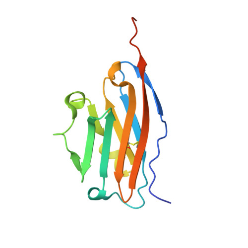
    9EO0

  • PubMed Abstract: 

    Therapeutic antibodies directed against either programmed cell death-1 protein (PD-1) or its ligand PD-L1 have demonstrated efficacy in the treatment of various cancers. In contrast with antibodies, small molecules have the potential for increased tissue penetration; better pharmacology; and therefore, improved antitumor activity. A series of nonsymmetric C2 inhibitors were synthesized and evaluated for PD-1/PD-L1 interaction inhibition. These compounds induced PD-L1 dimerization and effectively blocked PD-L1/PD-1 interaction in a homogeneous time-resolved fluorescence (HTRF) assay with most inhibitors exhibiting IC 50 values in the single-digit nM range and below. Their high inhibitory potency was also demonstrated in a cell-based coculture PD-1 signaling assay where 2 exhibited an EC 50 inhibitory activity of 21.8 nM, which approached that of the PD-L1 antibody durvalumab (EC 50 = 0.3-1.8 nM). Structural insight into how these inhibitors interact with PD-L1 was gained by using NMR and X-ray cocrystal structure studies. These data support further preclinical evaluation of these compounds as antibody alternatives.


  • Organizational Affiliation
    • Jagiellonian University, Doctoral School of Exact and Natural Sciences, prof. S. Łojasiewicza 11, 30-348 Krakow, Poland.

Macromolecule Content 

  • Total Structure Weight: 91.8 kDa 
  • Atom Count: 5,833 
  • Modeled Residue Count: 711 
  • Deposited Residue Count: 786 
  • Unique protein chains: 1

Macromolecules

Find similar proteins by:|  3D Structure
Entity ID: 1
MoleculeChains  Sequence LengthOrganismDetailsImage
Programmed cell death 1 ligand 1
A, B, C, D, E
A, B, C, D, E, F
131Homo sapiensMutation(s): 0 
Gene Names: CD274B7H1PDCD1L1PDCD1LG1PDL1
UniProt & NIH Common Fund Data Resources
Find proteins for Q9NZQ7 (Homo sapiens)
Explore Q9NZQ7 
Go to UniProtKB:  Q9NZQ7
PHAROS:  Q9NZQ7
GTEx:  ENSG00000120217 
Entity Groups
Sequence Clusters30% Identity50% Identity70% Identity90% Identity95% Identity100% Identity
UniProt GroupQ9NZQ7
Sequence Annotations
Expand
Reference Sequence

Experimental Data & Validation

Experimental Data

  • Method: X-RAY DIFFRACTION
  • Resolution: 2.50 Å
  • R-Value Free:  0.302 (Depositor), 0.302 (DCC) 
  • R-Value Work:  0.253 (Depositor), 0.253 (DCC) 
Space Group: P 1 21 1
Unit Cell:
Length ( Å )Angle ( ˚ )
a = 70.753α = 90
b = 75.405β = 97.249
c = 75.923γ = 90
Software Package:
Software NamePurpose
REFMACrefinement
REFMACrefinement
XDSdata reduction
Aimlessdata scaling
PHASERphasing

Structure Validation

View Full Validation Report


Ligand Structure Quality Assessment 


Entry History 

& Funding Information

Deposition Data


Funding OrganizationLocationGrant Number
National Center for Research and Development (Poland)PolandPOIR.01.01.01-00-0129/18

Revision History  (Full details and data files)

  • Version 1.0: 2024-06-19
    Type: Initial release
  • Version 1.1: 2024-07-03
    Changes: Database references
  • Version 1.2: 2024-10-16
    Changes: Structure summary