9EGA | pdb_00009ega

Crystal Structure of a CE20 carbohydrate acetylesterase from Pedobacter psychrotolerans (PpCE20_II), with ancillary domain


Experimental Data Snapshot

  • Method: X-RAY DIFFRACTION
  • Resolution: 1.35 Å
  • R-Value Free: 
    0.192 (Depositor), 0.201 (DCC) 
  • R-Value Work: 
    0.159 (Depositor), 0.167 (DCC) 
  • R-Value Observed: 
    0.166 (Depositor) 

Starting Model: experimental
View more details

wwPDB Validation 3D Report Full Report

Validation slider image for 9EGA

Ligand Structure Quality Assessment 


This is version 1.0 of the entry. See complete history

Literature

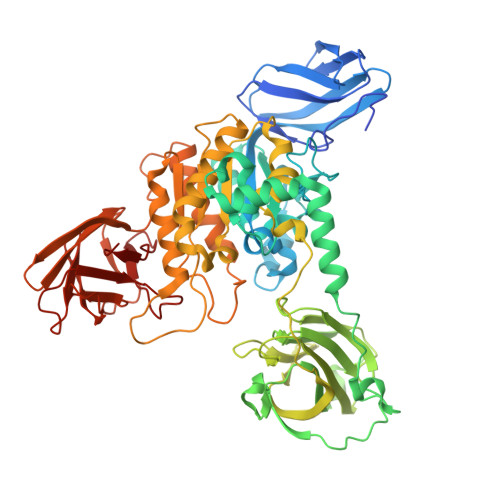
Insights into a water-mediated catalytic triad architecture in CE20 carbohydrate esterases.

Teune, M.Vieira, P.S.Dohler, T.Palm, G.J.Dutschei, T.Bartosik, D.Berndt, L.Persinoti, G.F.Maass, S.Becher, D.Schweder, T.Murakami, M.T.Lammers, M.Bornscheuer, U.T.

(2025) Nat Commun 16: 7034-7034

  • DOI: https://doi.org/10.1038/s41467-025-62387-5
  • Primary Citation Related Structures: 
    9EGA, 9H4U

  • PubMed Abstract: 

    Carbohydrate esterases modify polysaccharides by removing different ester moieties thereby affecting their physicochemical properties and their accessibility by glycoside hydrolases. We determined the full-length structures of two members (Fl8CE20_II and PpCE20_II) from the carbohydrate esterase family 20 (CE20) by X-ray crystallography that feature an ancillary domain, inserted into the catalytic SGNH-hydrolase domain. Detailed structural analysis identifies a so far undescribed catalytic triad architecture which lacks the typical aspartate for polarization of the histidine but instead reveals a precisely coordinated water molecule mediating contact between the His and Asp. This coordinated water in the Ser-His-(H 2 O-Asp/Asn) motif, as further confirmed by mutational studies and by determination of kinetic constants, is crucial for catalytic activity. We therefore term this active site architecture a water-mediated catalytic triad.


  • Organizational Affiliation
    • Department of Biotechnology & Enzyme Catalysis, Institute of Biochemistry, University Greifswald, Greifswald, Germany.

Macromolecule Content 

  • Total Structure Weight: 145.8 kDa 
  • Atom Count: 11,893 
  • Modeled Residue Count: 1,249 
  • Deposited Residue Count: 1,288 
  • Unique protein chains: 1

Macromolecules

Find similar proteins by:|  3D Structure
Entity ID: 1
MoleculeChains  Sequence LengthOrganismDetailsImage
Sialate O-acetylesterase
A, B
644Pedobacter psychrotoleransMutation(s): 0 
Gene Names: EV200_102244
UniProt
Find proteins for A0A4R2HIK5 (Pedobacter psychrotolerans)
Explore A0A4R2HIK5 
Go to UniProtKB:  A0A4R2HIK5
Entity Groups
Sequence Clusters30% Identity50% Identity70% Identity90% Identity95% Identity100% Identity
UniProt GroupA0A4R2HIK5
Sequence Annotations
Expand
Reference Sequence

Experimental Data & Validation

Experimental Data

  • Method: X-RAY DIFFRACTION
  • Resolution: 1.35 Å
  • R-Value Free:  0.192 (Depositor), 0.201 (DCC) 
  • R-Value Work:  0.159 (Depositor), 0.167 (DCC) 
  • R-Value Observed: 0.166 (Depositor) 
Space Group: P 1 21 1
Unit Cell:
Length ( Å )Angle ( ˚ )
a = 56.134α = 90
b = 125.135β = 104.71
c = 108.688γ = 90
Software Package:
Software NamePurpose
PHENIXrefinement
XDSdata scaling
XDSdata reduction
PHENIXphasing

Structure Validation

View Full Validation Report


Ligand Structure Quality Assessment 


Entry History 

& Funding Information

Deposition Data


Funding OrganizationLocationGrant Number
Sao Paulo Research Foundation (FAPESP)Brazil16/06509-0
Sao Paulo Research Foundation (FAPESP)Brazil15/26982-0

Revision History  (Full details and data files)

  • Version 1.0: 2025-08-13
    Type: Initial release