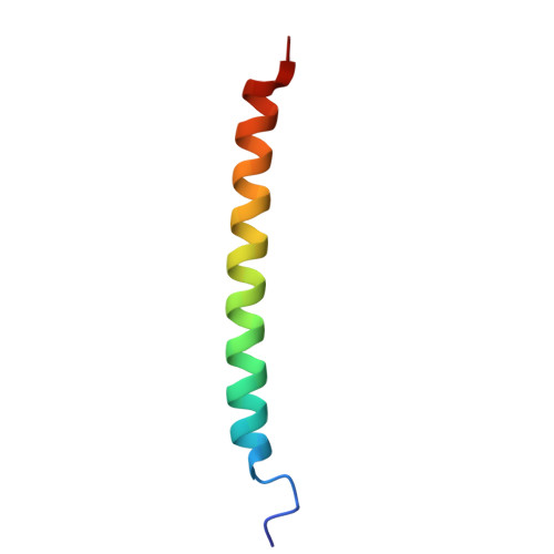
Functional unfolding of the integrin alpha X transmembrane helix.
Vu, H.N., Lee, M., Situ, A.J., An, W., Ley, K., Kim, C., Ulmer, T.S.(2025) Proc Natl Acad Sci U S A 122: e2507966122-e2507966122
- PubMed: 40956891 Search on PubMed
- DOI: https://doi.org/10.1073/pnas.2507966122
- Primary Citation Related Structures:
9ECL, 9ECM - PubMed Abstract:
In biological membranes, proteins face a fundamentally different environment than in water. To avoid untenable lipid contacts with polar backbone atoms, they use the continuous hydrogen bonding achieved by α-helices or β-barrels to traverse membranes. Here, we show that integrin αX, and by homology αM, undermine this paradigm by partially unfolding the N-terminal third of their transmembrane (TM) helix. Unfolding results in a dynamic, frayed helix that weakens the association with its partnering β2 subunit to lower the activation threshold of integrin αXβ2-mediated cell adhesion. The extent of unfolding depends on membrane geometry, thereby establishing a mechanism for sensing membrane properties. The combination of adhesive control with sensory capacity in integrin αXβ2 and αMβ2 may achieve membrane localization-dependent receptor activation in leukocyte phagocytosis. The unfolding of the αX TM helix arises from a high number of α-helix-destabilizing residues that TM helices in general approach but do not exceed. Accordingly, backbone dynamics of TM helices may disrupt hydrogen bonds, modulate protein function, and optimize TM helix rigidity.
- Department of Physiology and Neuroscience, Zilkha Neurogenetic Institute, Keck School of Medicine, University of Southern California, Los Angeles, CA 90033.
Organizational Affiliation: