9EB6 | pdb_00009eb6

Chicken YF1.7*1 presenting myristoylated peptide derived from tegument protein CIRC


Experimental Data Snapshot

  • Method: X-RAY DIFFRACTION
  • Resolution: 1.90 Å
  • R-Value Free: 
    0.221 (Depositor), 0.224 (DCC) 
  • R-Value Work: 
    0.171 (Depositor), 0.180 (DCC) 
  • R-Value Observed: 
    0.174 (Depositor) 

Starting Model: experimental
View more details

wwPDB Validation 3D Report Full Report

Validation slider image for 9EB6

Ligand Structure Quality Assessment 


This is version 1.0 of the entry. See complete history

Literature

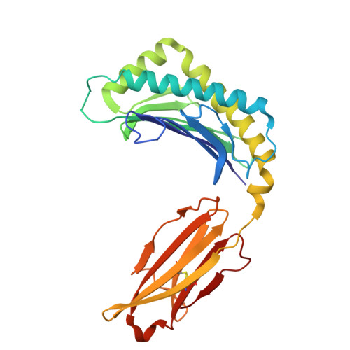
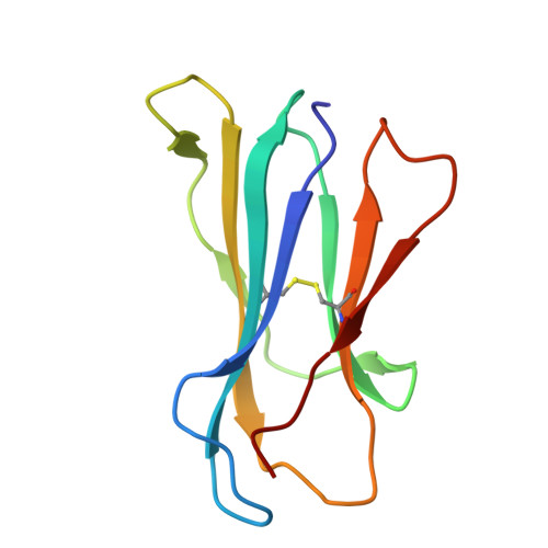
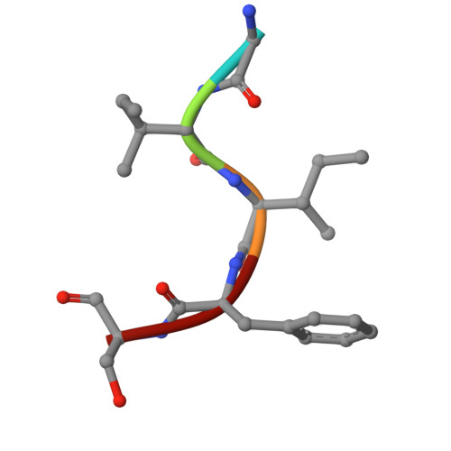
Molecular basis for presentation of N-myristoylated peptides by the chicken YF1∗7.1 molecule.

Khandokar, Y.Cheng, T.Y.Wang, C.J.H.Cao, T.P.Nagampalli, R.S.K.Sivaraman, K.K.Van Rhijn, I.Rossjohn, J.Moody, D.B.Nours, J.L.

(2025) J Biological Chem 301: 110253-110253

  • DOI: https://doi.org/10.1016/j.jbc.2025.110253
  • Primary Citation Related Structures: 
    9EB2, 9EB3, 9EB4, 9EB5, 9EB6

  • PubMed Abstract: 

    Major histocompatibility complex I (MHC-I) and MHC-I-like molecules play a central role in mediating immunity. Through their conservation across all taxa of jawed vertebrates, the MHC-I-like proteins have adapted to present non-peptidic antigens to distinct T cell populations. While our understanding of the structure-function relationship of MHC-I and MHC-I-like molecules in humans and mice is well established, the nature of the antigens presented by MHC-I- like molecules in "non-model" species remains unclear. Here, using a mammalian recombinant expression system combined with mass spectrometry approaches, we identified N-myristoylated peptides as endogenous ligands for the chicken MHC-I-like protein YF1∗7.1. Given the importance of N-myristoylation in viral pathogenesis, we determined the crystal structure of YF1∗7.1 in complex with two N-myristoylated peptides derived from Marek's disease virus (MDV), demonstrating the molecular basis that underpins the presentation of N-myristoylated peptides from MDV, a highly contagious and fatal viral neoplastic disease in chickens. Thus, the identified ligands are distinct from unmodified peptides found in classical MHC-I and -II as well as diverse amphipathic lipids captured by CD1 proteins. Collectively, our study lays the foundation for further molecular and functional characterization of YF1∗7.1 and more broadly of the role of the MHC-I encoded by the MHC-Y gene cluster in protection against highly contagious viral neoplastic diseases in chickens.


  • Organizational Affiliation
    • Infection and Immunity Program and Department of Biochemistry and Molecular Biology, Biomedicine Discovery Institute, Monash University, Clayton, Victoria, Australia.

Macromolecule Content 

  • Total Structure Weight: 43.26 kDa 
  • Atom Count: 3,255 
  • Modeled Residue Count: 369 
  • Deposited Residue Count: 373 
  • Unique protein chains: 3

Macromolecules

Find similar proteins by:|  3D Structure
Entity ID: 1
MoleculeChains  Sequence LengthOrganismDetailsImage
MHC Rfp-Y class I alpha chain270Gallus gallusMutation(s): 0 
Gene Names: YFV
UniProt
Find proteins for Q9BCW3 (Gallus gallus)
Explore Q9BCW3 
Go to UniProtKB:  Q9BCW3
Entity Groups
Sequence Clusters30% Identity50% Identity70% Identity90% Identity95% Identity100% Identity
UniProt GroupQ9BCW3
Sequence Annotations
Expand
Reference Sequence
Find similar proteins by:|  3D Structure
Entity ID: 2
MoleculeChains  Sequence LengthOrganismDetailsImage
Beta-2-microglobulin98Gallus gallusMutation(s): 0 
Gene Names: B2M
UniProt
Find proteins for P21611 (Gallus gallus)
Explore P21611 
Go to UniProtKB:  P21611
Entity Groups
Sequence Clusters30% Identity50% Identity70% Identity90% Identity95% Identity100% Identity
UniProt GroupP21611
Sequence Annotations
Expand
Reference Sequence
Find similar proteins by:  Sequence   |   3D Structure  
Entity ID: 3
MoleculeChains  Sequence LengthOrganismDetailsImage
Peptide derived from tegument protein CIRC5Marek's disease herpesvirus type 1 strain MD5Mutation(s): 0 

Small Molecules

Ligands 5 Unique
IDChains Name / Formula / InChI Key2D Diagram3D Interactions
MYR
(Subject of Investigation/LOI)

Query on MYR


Download:Ideal Coordinates CCD File
K [auth C]MYRISTIC ACID
C14 H28 O2
TUNFSRHWOTWDNC-UHFFFAOYSA-N
PGE

Query on PGE


Download:Ideal Coordinates CCD File
D [auth A]TRIETHYLENE GLYCOL
C6 H14 O4
ZIBGPFATKBEMQZ-UHFFFAOYSA-N
GOL

Query on GOL


Download:Ideal Coordinates CCD File
E [auth A]GLYCEROL
C3 H8 O3
PEDCQBHIVMGVHV-UHFFFAOYSA-N
CL

Query on CL


Download:Ideal Coordinates CCD File
F [auth A],
G [auth A],
H [auth A]
CHLORIDE ION
Cl
VEXZGXHMUGYJMC-UHFFFAOYSA-M
NA

Query on NA


Download:Ideal Coordinates CCD File
I [auth A],
J [auth A]
SODIUM ION
Na
FKNQFGJONOIPTF-UHFFFAOYSA-N

Experimental Data & Validation

Experimental Data

  • Method: X-RAY DIFFRACTION
  • Resolution: 1.90 Å
  • R-Value Free:  0.221 (Depositor), 0.224 (DCC) 
  • R-Value Work:  0.171 (Depositor), 0.180 (DCC) 
  • R-Value Observed: 0.174 (Depositor) 
Space Group: I 1 2 1
Unit Cell:
Length ( Å )Angle ( ˚ )
a = 55.289α = 90
b = 55.41β = 97.92
c = 137.904γ = 90
Software Package:
Software NamePurpose
MOSFLMdata reduction
Aimlessdata scaling
PHASERphasing
REFMACrefinement
PHENIXrefinement

Structure Validation

View Full Validation Report


Ligand Structure Quality Assessment 


Entry History 

& Funding Information

Deposition Data


Funding OrganizationLocationGrant Number
National Health and Medical Research Council (NHMRC, Australia)Australia2008616
Monash University/ARC Centre of Excellence in Advanced Molecular Imaging AllianceAustraliaCE140100011

Revision History  (Full details and data files)

  • Version 1.0: 2025-07-23
    Type: Initial release