9E6Y | pdb_00009e6y

Structure of CD112 (Nectin-2) domain 1 bound to CD112R (PVRIG)


Experimental Data Snapshot

  • Method: X-RAY DIFFRACTION
  • Resolution: 2.20 Å
  • R-Value Free: 
    0.245 (Depositor), 0.246 (DCC) 
  • R-Value Work: 
    0.208 (Depositor), 0.207 (DCC) 
  • R-Value Observed: 
    0.210 (Depositor) 

Starting Models: experimental, in silico
View more details

wwPDB Validation 3D Report Full Report

Validation slider image for 9E6Y

Ligand Structure Quality Assessment 


This is version 1.1 of the entry. See complete history

Literature

Structure-guided engineering of CD112 receptor variants for optimized immunotherapy.

Singh, S.Julia, E.Kalita, P.Mason, C.Ming, Q.Lee-Sam, A.Gordon, S.Buitrago, M.E.Leung, D.W.Hwu, P.Luca, V.C.

(2025) Mol Ther 33: 3590-3604

  • DOI: https://doi.org/10.1016/j.ymthe.2025.04.032
  • Primary Citation Related Structures: 
    9E6Y

  • PubMed Abstract: 

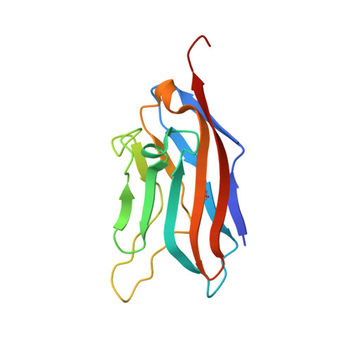
    The immune checkpoint protein, CD112 receptor (CD112R, also known as PVRIG), suppresses T and NK cell activation upon binding to tumor-expressed CD112 (Nectin-2) ligands. Here, we determine the structure of the CD112-CD112R complex and use it to guide the engineering of multiple CD112-targeting immunotherapy candidates. The 2.2 Å-resolution crystal structure reveals an antiparallel, lock-and-key binding mode in which CD112R disrupts CD112 homodimerization. Structural analysis informed directed evolution campaigns focused on remodeling the CD112-CD112R interface, resulting in the isolation of CD112R mutants with greatly increased expression and CD112-binding affinity. The highest-affinity variant, CD112R IVE , potently inhibits CD112-CD112R interactions when utilized as a soluble CD112 trap. Furthermore, incorporating CD112R variants into chimeric antigen receptors (CARs) and T cell engagers (TCEs) leads to more robust T cell activation and killing of CD112 + triple-negative breast cancer (TNBC) cells compared to wild-type CD112R. This strategy demonstrates how structural insights can be leveraged to efficiently generate panels of "affinity-tuned" biologics for immunotherapy.


  • Organizational Affiliation
    • Department of Immunology, H. Lee Moffitt Cancer Center and Research Institute, Tampa, FL, 33612, USA; Cancer Biology Ph.D. Program, University of South Florida, Tampa, FL 33612, USA.

Macromolecule Content 

  • Total Structure Weight: 29.25 kDa 
  • Atom Count: 1,973 
  • Modeled Residue Count: 238 
  • Deposited Residue Count: 265 
  • Unique protein chains: 2

Macromolecules

Find similar proteins by:|  3D Structure
Entity ID: 1
MoleculeChains  Sequence LengthOrganismDetailsImage
Nectin-2A [auth D]132Homo sapiensMutation(s): 0 
Gene Names: NECTIN2HVEBPRR2PVRL2
UniProt & NIH Common Fund Data Resources
Find proteins for Q92692 (Homo sapiens)
Explore Q92692 
Go to UniProtKB:  Q92692
PHAROS:  Q92692
GTEx:  ENSG00000130202 
Entity Groups
Sequence Clusters30% Identity50% Identity70% Identity90% Identity95% Identity100% Identity
UniProt GroupQ92692
Sequence Annotations
Expand
Reference Sequence
Find similar proteins by:|  3D Structure
Entity ID: 2
MoleculeChains  Sequence LengthOrganismDetailsImage
Transmembrane protein PVRIGB [auth A]133Homo sapiensMutation(s): 0 
Gene Names: PVRIGC7orf15
UniProt & NIH Common Fund Data Resources
Find proteins for Q6DKI7 (Homo sapiens)
Explore Q6DKI7 
Go to UniProtKB:  Q6DKI7
GTEx:  ENSG00000213413 
Entity Groups
Sequence Clusters30% Identity50% Identity70% Identity90% Identity95% Identity100% Identity
UniProt GroupQ6DKI7
Glycosylation
Glycosylation Sites: 1Go to GlyGen: Q6DKI7-1
Sequence Annotations
Expand
Reference Sequence

Small Molecules

Ligands 2 Unique
IDChains Name / Formula / InChI Key2D Diagram3D Interactions
NAG
(Subject of Investigation/LOI)

Query on NAG


Download:Ideal Coordinates CCD File
I [auth A]2-acetamido-2-deoxy-beta-D-glucopyranose
C8 H15 N O6
OVRNDRQMDRJTHS-FMDGEEDCSA-N
EDO
(Subject of Investigation/LOI)

Query on EDO


Download:Ideal Coordinates CCD File
C [auth D]
D
E [auth D]
F [auth D]
G [auth D]
C [auth D],
D,
E [auth D],
F [auth D],
G [auth D],
H [auth D],
J [auth A],
K [auth A],
L [auth A],
M [auth A],
N [auth A]
1,2-ETHANEDIOL
C2 H6 O2
LYCAIKOWRPUZTN-UHFFFAOYSA-N

Experimental Data & Validation

Experimental Data

  • Method: X-RAY DIFFRACTION
  • Resolution: 2.20 Å
  • R-Value Free:  0.245 (Depositor), 0.246 (DCC) 
  • R-Value Work:  0.208 (Depositor), 0.207 (DCC) 
  • R-Value Observed: 0.210 (Depositor) 
Space Group: P 43 21 2
Unit Cell:
Length ( Å )Angle ( ˚ )
a = 79.65α = 90
b = 79.65β = 90
c = 81.38γ = 90
Software Package:
Software NamePurpose
PHENIXrefinement
PHENIXrefinement
XDSdata reduction
XDSdata scaling
PHENIXphasing

Structure Validation

View Full Validation Report


Ligand Structure Quality Assessment 


Entry History 

& Funding Information

Deposition Data


Funding OrganizationLocationGrant Number
National Institutes of Health/National Institute of General Medical Sciences (NIH/NIGMS)United StatesGM133482-01

Revision History  (Full details and data files)

  • Version 1.0: 2025-05-07
    Type: Initial release
  • Version 1.1: 2025-08-20
    Changes: Database references