9E0V | pdb_00009e0v

GSDMD bound to a peptide


Experimental Data Snapshot

  • Method: X-RAY DIFFRACTION
  • Resolution: 1.64 Å
  • R-Value Free: 
    0.212 (Depositor), 0.209 (DCC) 
  • R-Value Work: 
    0.200 (Depositor), 0.192 (DCC) 
  • R-Value Observed: 
    0.201 (Depositor) 

Starting Model: experimental
View more details

wwPDB Validation 3D Report Full Report

Validation slider image for 9E0V

This is version 1.3 of the entry. See complete history

Literature

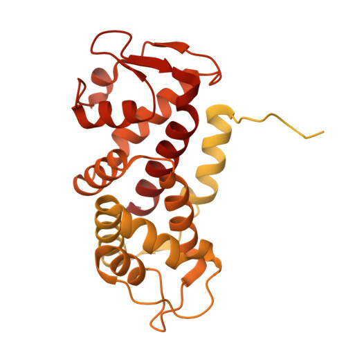
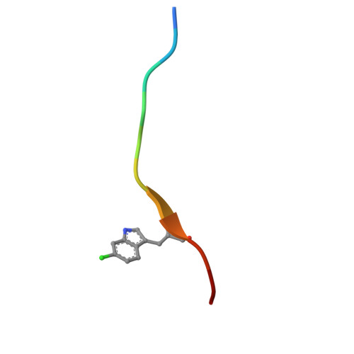
Peptide binding at the gasdermin D exosite reveals the structural basis for targeting the site.

Wang, R.Ho, T.Ogawa, A.Widjaja, K.Brown, Z.Yang, S.Huo, P.Gregory, M.C.Singh, A.Perumal, S.Lin, S.Cheng, A.C.Garrenton, L.S.Mu, J.Ogawa, A.Chrencik, J.E.

(2026) Acta Crystallogr D Struct Biol 82: 615-625

  • DOI: https://doi.org/10.1107/S205979832600344X
  • Primary Citation Related Structures: 
    9E0V

  • PubMed Abstract: 

    Gasdermin D (GSDMD) has been identified as a critical component of the inflammasome, an important intracellular signaling multi-protein complex. Abnormal activation of GSDMD has been linked to a variety of inflammatory diseases, including non-alcoholic steatohepatitis, inflammatory bowel disease and COVID-19, and has been linked to potential pathogenesis of septic shock. While small molecules have been identified to inhibit N-terminal domain (NTD) pore formation and membrane translocation, further clinical application of these molecules is currently limited due to narrow specificity. Here, we report a peptide from a structure-based computational screen that selectively inhibits caspase-dependent cleavage of GSDMD. We have determined the structure of the GSDMD-peptide complex, indicating that peptide binding is facilitated by negatively charged residues from the peptide and the hydrophobic exosite of the protein, inhibiting caspase binding and activation. The experimental data suggest the potential to target the exosite for therapeutic intervention.


  • Organizational Affiliation
    • Protein and Structural Chemistry, Merck & Co., Inc, 213 East Grand Avenue, South San Francisco, CA 94080, USA.

Macromolecule Content 

  • Total Structure Weight: 65.42 kDa 
  • Atom Count: 1,759 
  • Modeled Residue Count: 215 
  • Deposited Residue Count: 601 
  • Unique protein chains: 2

Macromolecules

Find similar proteins by:|  3D Structure
Entity ID: 1
MoleculeChains  Sequence LengthOrganismDetailsImage
Maltose/maltodextrin-binding periplasmic protein, Gasdermin-D, C-terminal chimera590Homo sapiensMutation(s): 0 
Gene Names: malEb4034JW3994GSDMDDFNA5LGSDMDC1FKSG10
UniProt & NIH Common Fund Data Resources
Find proteins for P57764 (Homo sapiens)
Explore P57764 
Go to UniProtKB:  P57764
PHAROS:  P57764
GTEx:  ENSG00000104518 
Entity Groups
Sequence Clusters30% Identity50% Identity70% Identity90% Identity95% Identity100% Identity
UniProt GroupP57764
Sequence Annotations
Expand
Reference Sequence
Find similar proteins by:  Sequence   |   3D Structure  
Entity ID: 2
MoleculeChains  Sequence LengthOrganismDetailsImage
GLY-CYS-ILE-LYS-LYS-ALA-VAL-6CW-PHE-LYS-CYS11Homo sapiensMutation(s): 0 

Small Molecules

Modified Residues  1 Unique
IDChains TypeFormula2D DiagramParent
6CW
Query on 6CW
B
L-PEPTIDE LINKINGC11 H11 Cl N2 O2TRP

Experimental Data & Validation

Experimental Data

  • Method: X-RAY DIFFRACTION
  • Resolution: 1.64 Å
  • R-Value Free:  0.212 (Depositor), 0.209 (DCC) 
  • R-Value Work:  0.200 (Depositor), 0.192 (DCC) 
  • R-Value Observed: 0.201 (Depositor) 
Space Group: P 43 21 2
Unit Cell:
Length ( Å )Angle ( ˚ )
a = 47.575α = 90
b = 47.575β = 90
c = 194.717γ = 90
Software Package:
Software NamePurpose
BUSTERrefinement
pointlessdata scaling
XDSdata reduction
PHENIXphasing

Structure Validation

View Full Validation Report


Entry History 

& Funding Information

Deposition Data

  • Released Date: 2025-10-22 
  • Deposition Author(s): Wang, R.

Funding OrganizationLocationGrant Number
Not funded--

Revision History  (Full details and data files)

  • Version 1.0: 2025-10-22
    Type: Initial release
  • Version 1.1: 2026-02-11
    Changes: Data collection
  • Version 1.2: 2026-05-27
    Changes: Database references
  • Version 1.3: 2026-06-10
    Changes: Database references