9DZH | pdb_00009dzh

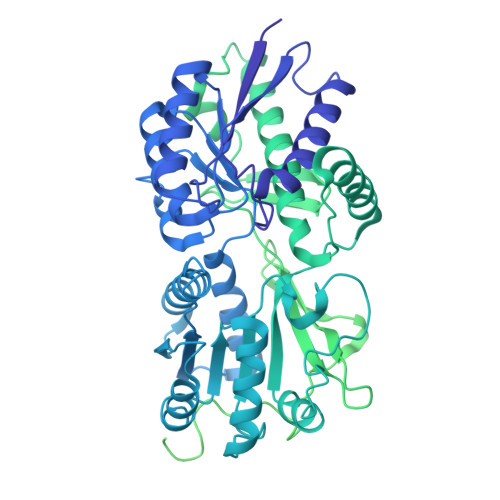
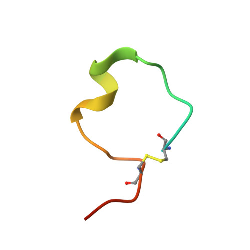
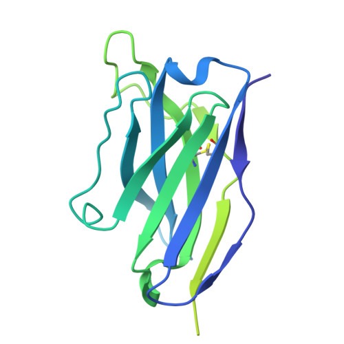
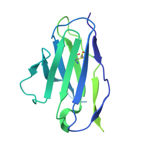
Human GC-A bound to ANP and REGN5308


Experimental Data Snapshot

  • Method: ELECTRON MICROSCOPY
  • Resolution: 3.00 Å
  • Aggregation State: PARTICLE 
  • Reconstruction Method: SINGLE PARTICLE 

wwPDB Validation 3D Report Full Report

Validation slider image for 9DZH

This is version 1.1 of the entry. See complete history. 

Literature

Structural insights into single-pass transmembrane receptor GC-A activation by distinct antihypertensive antibodies.

Liu, S., Manzo, O., Wang, J., Zhu, L., Xiao, F., Su, Y.C., Kurre, D., Liu, W., Miao, Y., Di Lorenzo, A., Huang, X.Y.

(2026) Nat Commun 17

  • DOI: https://doi.org/10.1038/s41467-026-71594-7
  • Primary Citation Related Structures: 
    9DZF, 9DZG, 9DZH, 9DZJ, 9DZK

  • PubMed Abstract: 

    The single-pass transmembrane receptor guanylyl cyclase A (GC-A), also known as natriuretic peptide receptor A (NPR-A) or NPR1, regulates blood pressure through vasodilation and natriuresis, making it a promising therapeutic target for hypertension and heart failure. We describe two monoclonal antibodies, XX16 and REGN5308, that differentially activate GC-A. Using cryo-electron microscopy and molecular dynamics simulations, we reveal that XX16 stabilizes GC-A in an active conformation even without its ligand ANP, whereas REGN5308 requires ANP to fully promote receptor activation. Both antibodies increase ANP binding affinity to GC-A and enhance GC-A-mediated cGMP signaling, although XX16 exerts a stronger stabilizing influence on ATP and GTP binding. In a mouse model of obesity-induced hypertension, XX16 treatment significantly reduces blood pressure, underscoring its therapeutic potential. These findings outline the structural and functional basis of GC-A activation by antibody positive allosteric modulators, offering strategies for durable antihypertensive therapies and improved management of cardiovascular diseases.


  • Organizational Affiliation: 
    • Department of Biochemistry and Biophysics, Weill Medical College of Cornell University, New York, NY, USA.

Macromolecule Content 

  • Total Structure Weight: 285.47 kDa 
  • Atom Count: 8,425 
  • Modeled Residue Count: 1,103 
  • Deposited Residue Count: 2,530 
  • Unique protein chains: 4

Macromolecules

Find similar proteins by:|  3D Structure
Entity ID: 1
MoleculeChains  Sequence LengthOrganismDetailsImage
Atrial natriuretic peptide receptor 1
A, B
1,031Homo sapiensMutation(s): 0 
Gene Names: NPR1, ANPRA
EC: 4.6.1.2
UniProt & NIH Common Fund Data Resources
Find proteins for P16066 (Homo sapiens)
Explore P16066 
Go to UniProtKB:  P16066
PHAROS:  P16066
GTEx:  ENSG00000169418 
Entity Groups
Sequence Clusters30% Identity50% Identity70% Identity90% Identity95% Identity100% Identity
UniProt GroupP16066
Glycosylation
Glycosylation Sites: 4Go to GlyGen: P16066-1
Sequence Annotations
Expand
Reference Sequence
Find similar proteins by:|  3D Structure
Entity ID: 2
MoleculeChains  Sequence LengthOrganismDetailsImage
Atrial natriuretic peptide28Homo sapiensMutation(s): 0 
Gene Names: NPPA, ANP, PND
UniProt & NIH Common Fund Data Resources
Find proteins for P01160 (Homo sapiens)
Explore P01160 
Go to UniProtKB:  P01160
PHAROS:  P01160
GTEx:  ENSG00000175206 
Entity Groups
Sequence Clusters30% Identity50% Identity70% Identity90% Identity95% Identity100% Identity
UniProt GroupP01160
Sequence Annotations
Expand
Reference Sequence
Find similar proteins by:|  3D Structure
Entity ID: 3
MoleculeChains  Sequence LengthOrganismDetailsImage
REGN5308 - Heavy chainD [auth H]227Mus musculusMutation(s): 0 
Entity Groups
Sequence Clusters30% Identity50% Identity70% Identity90% Identity95% Identity100% Identity
Sequence Annotations
Expand
Reference Sequence
Find similar proteins by:|  3D Structure
Entity ID: 4
MoleculeChains  Sequence LengthOrganismDetailsImage
REGN5308 - Light chainE [auth L]213Mus musculusMutation(s): 0 
Entity Groups
Sequence Clusters30% Identity50% Identity70% Identity90% Identity95% Identity100% Identity
Sequence Annotations
Expand
Reference Sequence

Oligosaccharides

Help  
Entity ID: 5
MoleculeChains Length2D Diagram GlycosylationD Interactions
2-acetamido-2-deoxy-beta-D-glucopyranose-(1-4)-2-acetamido-2-deoxy-beta-D-glucopyranoseF [auth D],
H [auth F],
J [auth I]
2N-Glycosylation
Glycosylation Resources
GlyTouCan: G42666HT
GlyCosmos: G42666HT
GlyGen: G42666HT
Entity ID: 6
MoleculeChains Length2D Diagram GlycosylationD Interactions
beta-D-mannopyranose-(1-4)-2-acetamido-2-deoxy-beta-D-glucopyranose-(1-4)-2-acetamido-2-deoxy-beta-D-glucopyranoseG [auth E],
I [auth G]
3N-Glycosylation
Glycosylation Resources
GlyTouCan: G15407YE
GlyCosmos: G15407YE
GlyGen: G15407YE

Experimental Data & Validation

Experimental Data

  • Method: ELECTRON MICROSCOPY
  • Resolution: 3.00 Å
  • Aggregation State: PARTICLE 
  • Reconstruction Method: SINGLE PARTICLE 
EM Software:
TaskSoftware PackageVersion
MODEL REFINEMENTPHENIX1.20.1_4487:

Structure Validation

View Full Validation Report


Entry History 

& Funding Information

Deposition Data


Funding OrganizationLocationGrant Number
National Institutes of Health/National Institute of General Medical Sciences (NIH/NIGMS)United States--

Revision History  (Full details and data files)

  • Version 1.0: 2026-01-21
    Type: Initial release
  • Version 1.1: 2026-08-05
    Changes: Data collection, Database references