


Crystal Structure of Human DAPK1 Catalytic Subunit Complexed with Compound SRM-07-081a
Minasov, G., Winsor, J., Roy, S.M., Watterson, D.M., Shuvalova, L.To be published.
Experimental Data Snapshot
Starting Model: experimental
View more details
Entity ID: 1 | |||||
|---|---|---|---|---|---|
| Molecule | Chains | Sequence Length | Organism | Details | Image |
| Death-associated protein kinase 1 | 294 | Homo sapiens | Mutation(s): 0 Gene Names: DAPK1, DAPK EC: 2.7.11.1 | 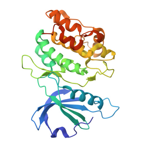 | |
UniProt & NIH Common Fund Data Resources | |||||
PHAROS: P53355 GTEx: ENSG00000196730 | |||||
Entity Groups | |||||
| Sequence Clusters | 30% Identity50% Identity70% Identity90% Identity95% Identity100% Identity | ||||
| UniProt Group | P53355 | ||||
Sequence AnnotationsExpand | |||||
Reference Sequence | |||||
| Ligands 3 Unique | |||||
|---|---|---|---|---|---|
| ID | Chains | Name / Formula / InChI Key | 2D Diagram | 3D Interactions | |
| A1BB6 (Subject of Investigation/LOI) Download:Ideal Coordinates CCD File | B [auth A] | 3-chloro-6-(4-methylpiperazin-1-yl)-4-(pyridin-4-yl)pyridazine C14 H16 Cl N5 GEGHOUWQCHWMKH-UHFFFAOYSA-N |  | ||
| SO4 Download:Ideal Coordinates CCD File | D [auth A], E [auth A], F [auth A], G [auth A], H [auth A] | SULFATE ION O4 S QAOWNCQODCNURD-UHFFFAOYSA-L |  | ||
| BO3 Download:Ideal Coordinates CCD File | C [auth A] | BORIC ACID B H3 O3 KGBXLFKZBHKPEV-UHFFFAOYSA-N |  | ||
| Length ( Å ) | Angle ( ˚ ) |
|---|---|
| a = 47.01 | α = 90 |
| b = 62.684 | β = 90 |
| c = 88.462 | γ = 90 |
| Software Name | Purpose |
|---|---|
| REFMAC | refinement |
| HKL-3000 | data reduction |
| HKL-3000 | data scaling |
| PHASER | phasing |
| Funding Organization | Location | Grant Number |
|---|---|---|
| National Institutes of Health/National Institute on Aging (NIH/NIA) | United States | AG066722 |


