


Crystal structure of ADP-ribose diphosphatase from Klebsiella pneumoniae (DTP bound, P21 form)
Liu, L., Lovell, S., Buchko, G.W., Battaile, K.P.To be published.
Experimental Data Snapshot
Starting Model: experimental
View more details
Entity ID: 1 | |||||
|---|---|---|---|---|---|
| Molecule | Chains | Sequence Length | Organism | Details | Image |
| ADP-ribose pyrophosphatase | 218 | Klebsiella pneumoniae | Mutation(s): 0 Gene Names: KPHS_45750 EC: 3.6.1.13 | 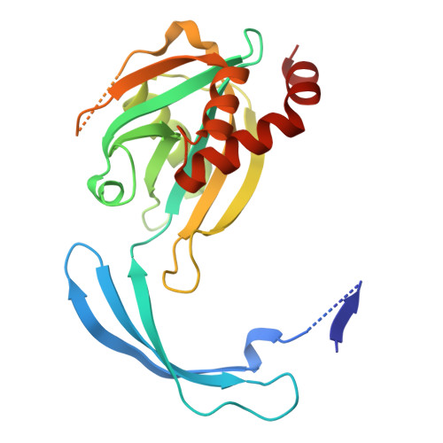 | |
UniProt | |||||
Entity Groups | |||||
| Sequence Clusters | 30% Identity50% Identity70% Identity90% Identity95% Identity100% Identity | ||||
| UniProt Group | B5XU49 | ||||
Sequence AnnotationsExpand | |||||
Reference Sequence | |||||
| Ligands 2 Unique | |||||
|---|---|---|---|---|---|
| ID | Chains | Name / Formula / InChI Key | 2D Diagram | 3D Interactions | |
| DTP (Subject of Investigation/LOI) Download:Ideal Coordinates CCD File | I [auth A] J [auth B] L [auth C] M [auth C] O [auth E] | 2'-DEOXYADENOSINE 5'-TRIPHOSPHATE C10 H16 N5 O12 P3 SUYVUBYJARFZHO-RRKCRQDMSA-N |  | ||
| MG (Subject of Investigation/LOI) Download:Ideal Coordinates CCD File | K [auth B] N [auth C] Q [auth E] R [auth F] S [auth F] | MAGNESIUM ION Mg JLVVSXFLKOJNIY-UHFFFAOYSA-N |  | ||
| Length ( Å ) | Angle ( ˚ ) |
|---|---|
| a = 93.034 | α = 90 |
| b = 80.131 | β = 90.13 |
| c = 116.189 | γ = 90 |
| Software Name | Purpose |
|---|---|
| PHENIX | refinement |
| Aimless | data scaling |
| XDS | data reduction |
| PHASER | phasing |
| Funding Organization | Location | Grant Number |
|---|---|---|
| National Institutes of Health/National Institute Of Allergy and Infectious Diseases (NIH/NIAID) | United States | 75N93022C00036 |
| National Institutes of Health/Office of the Director | United States | S10OD030394 |


