


Crystal structure of L-asparaginase from Streptococcus pneumoniae TIGR4
Gade, P., Endres, M., Babnigg, G., Joachimiak, A.To be published.
Experimental Data Snapshot
Starting Model: in silico
View more details
wwPDB Validation 3D Report Full Report

Entity ID: 1 | |||||
|---|---|---|---|---|---|
| Molecule | Chains | Sequence Length | Organism | Details | Image |
| Asparaginase | 320 | Streptococcus pneumoniae | Mutation(s): 0 Gene Names: ansB, A5N45_01685, AZJ70_05345, AZK02_05425, ERS019316_00818, ERS019420_01259, ERS021218_01180, GM536_02705, GM543_02955, GM545_09810... EC: 3.5.1.1 | 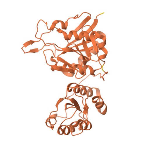 | |
UniProt | |||||
Find proteins for A0A0H2ZNF4 (Streptococcus pneumoniae serotype 2 (strain D39 / NCTC 7466)) Explore A0A0H2ZNF4 Go to UniProtKB: A0A0H2ZNF4 | |||||
Entity Groups | |||||
| Sequence Clusters | 30% Identity50% Identity70% Identity90% Identity95% Identity100% Identity | ||||
| UniProt Group | A0A0H2ZNF4 | ||||
Sequence AnnotationsExpand | |||||
Reference Sequence | |||||
| Ligands 2 Unique | |||||
|---|---|---|---|---|---|
| ID | Chains | Name / Formula / InChI Key | 2D Diagram | 3D Interactions | |
| SO4 Download:Ideal Coordinates CCD File | C [auth A], D [auth A], E [auth A], F [auth A] | SULFATE ION O4 S QAOWNCQODCNURD-UHFFFAOYSA-L |  | ||
| EDO Download:Ideal Coordinates CCD File | B [auth A], G [auth A] | 1,2-ETHANEDIOL C2 H6 O2 LYCAIKOWRPUZTN-UHFFFAOYSA-N |  | ||
| Length ( Å ) | Angle ( ˚ ) |
|---|---|
| a = 54.369 | α = 90 |
| b = 74.016 | β = 90 |
| c = 132.655 | γ = 90 |
| Software Name | Purpose |
|---|---|
| PHENIX | refinement |
| HKL-3000 | data reduction |
| HKL-3000 | data scaling |
| MOLREP | phasing |
| Funding Organization | Location | Grant Number |
|---|---|---|
| National Institutes of Health/National Institute Of Allergy and Infectious Diseases (NIH/NIAID) | United States | -- |


