9D65

Nitrile hydratase BR52A mutant


Experimental Data Snapshot

  • Method: X-RAY DIFFRACTION
  • Resolution: 1.45 Å
  • R-Value Free: 0.279 
  • R-Value Work: 0.240 
  • R-Value Observed: 0.242 

Starting Model: experimental
View more details

wwPDB Validation   3D Report Full Report


This is version 1.0 of the entry. See complete history


Literature

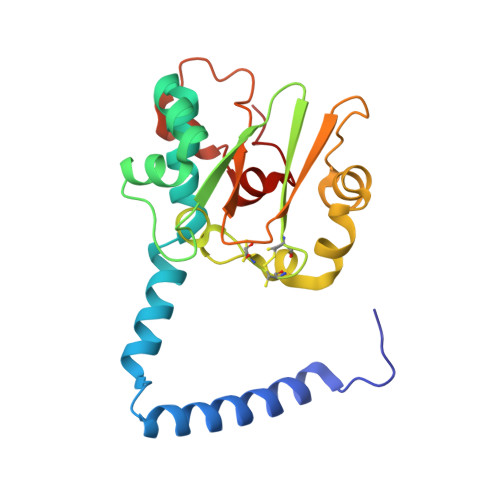
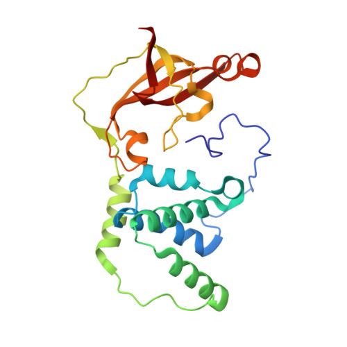
Role of second-sphere arginine residues in metal binding and metallocentre assembly in nitrile hydratases.

Miller, C.Huntoon, D.Kaley, N.Ogutu, I.Fiedler, A.T.Bennett, B.Liu, D.Holz, R.

(2024) J Inorg Biochem 256: 112565

  • DOI: https://doi.org/10.1016/j.jinorgbio.2024.112565
  • Primary Citation of Related Structures:  
    9D65, 9D6J, 9D6M

  • PubMed Abstract: 

    Two conserved second-sphere βArg (R) residues in nitrile hydratases (NHase), that form hydrogen bonds with the catalytically essential sulfenic and sulfinic acid ligands, were mutated to Lys and Ala residues in the Co-type NHase from Pseudonocardia thermophila JCM 3095 (PtNHase) and the Fe-type NHase from Rhodococcus equi TG328-2 (ReNHase). Only five of the eight mutants (PtNHase βR52A, βR52K, βR157A, βR157K and ReNHase βR61A) were successfully expressed and purified. Apart from the PtNHase βR52A mutant that exhibited no detectable activity, the k cat values obtained for the PtNHase and ReNHase βR mutant enzymes were between 1.8 and 12.4 s -1 amounting to <1% of the k cat values observed for WT enzymes. The metal content of each mutant was also significantly decreased with occupancies ranging from ∼10 to ∼40%. UV-Vis spectra coupled with EPR data obtained on the ReNHase mutant enzyme, suggest a decrease in the Lewis acidity of the active site metal ion. X-ray crystal structures of the four PtNHase βR mutant enzymes confirmed the mutation and the low active site metal content, while also providing insight into the active site hydrogen bonding network. Finally, DFT calculations suggest that the equatorial sulfenic acid ligand, which has been shown to be the catalytic nucleophile, is protonated in the mutant enzyme. Taken together, these data confirm the necessity of the conserved second-sphere βR residues in the proposed subunit swapping process and post-translational modification of the α-subunit in the α activator complex, along with stabilizing the catalytic sulfenic acid in its anionic form.


  • Organizational Affiliation

    Department of Chemistry and Geochemistry, Colorado School of Mines, Golden, CO 80401, USA.


Macromolecules
Find similar proteins by:  (by identity cutoff)  |  3D Structure
Entity ID: 1
MoleculeChains Sequence LengthOrganismDetailsImage
Cobalt-containing nitrile hydratase subunit alpha204Pseudonocardia thermophilaMutation(s): 0 
EC: 4.2.1.84
UniProt
Find proteins for Q7SID2 (Pseudonocardia thermophila)
Explore Q7SID2 
Go to UniProtKB:  Q7SID2
Entity Groups  
Sequence Clusters30% Identity50% Identity70% Identity90% Identity95% Identity100% Identity
UniProt GroupQ7SID2
Sequence Annotations
Expand
  • Reference Sequence
Find similar proteins by:  (by identity cutoff)  |  3D Structure
Entity ID: 2
MoleculeChains Sequence LengthOrganismDetailsImage
Cobalt-containing nitrile hydratase subunit beta228Pseudonocardia thermophilaMutation(s): 1 
EC: 4.2.1.84
UniProt
Find proteins for Q7SID3 (Pseudonocardia thermophila)
Explore Q7SID3 
Go to UniProtKB:  Q7SID3
Entity Groups  
Sequence Clusters30% Identity50% Identity70% Identity90% Identity95% Identity100% Identity
UniProt GroupQ7SID3
Sequence Annotations
Expand
  • Reference Sequence
Small Molecules
Modified Residues  1 Unique
IDChains TypeFormula2D DiagramParent
CSO
Query on CSO
A
L-PEPTIDE LINKINGC3 H7 N O3 SCYS
Experimental Data & Validation

Experimental Data

  • Method: X-RAY DIFFRACTION
  • Resolution: 1.45 Å
  • R-Value Free: 0.279 
  • R-Value Work: 0.240 
  • R-Value Observed: 0.242 
  • Space Group: P 32 2 1
Unit Cell:
Length ( Å )Angle ( ˚ )
a = 65.964α = 90
b = 65.964β = 90
c = 186.4γ = 120
Software Package:
Software NamePurpose
PHENIXrefinement
SCALAdata scaling
PHASERphasing
Adxvdata reduction

Structure Validation

View Full Validation Report


Entry History & Funding Information

Deposition Data


Funding OrganizationLocationGrant Number
National Science Foundation (NSF, United States)United States--

Revision History  (Full details and data files)

  • Version 1.0: 2024-08-28
    Type: Initial release