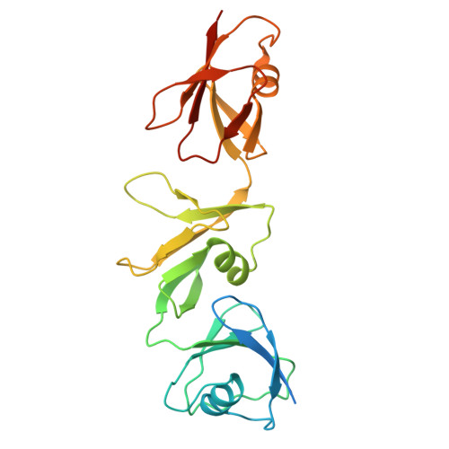
9D5A | pdb_00009d5a

Structure of Citrobacter multi-ubiquitin protein, local refinement of one full-length protomer


Experimental Data Snapshot

  • Method: ELECTRON MICROSCOPY
  • Resolution: 2.73 Å
  • Aggregation State: FILAMENT 
  • Reconstruction Method: SINGLE PARTICLE 

wwPDB Validation 3D Report Full Report

Validation slider image for 9D5A

This is version 1.2 of the entry. See complete history

Literature

Structural diversity and oligomerization of bacterial ubiquitin-like proteins.

Gong, M.Ye, Q.Gu, Y.Chambers, L.R.Bobkov, A.A.Arakawa, N.K.Matyszewski, M.Corbett, K.D.

(2025) Structure 33: 1016-1026.e4

  • DOI: https://doi.org/10.1016/j.str.2025.03.011
  • Primary Citation Related Structures: 
    8U38, 9CD2, 9D59, 9D5A, 9D5B

  • PubMed Abstract: 

    Bacteria possess a variety of operons with homology to eukaryotic ubiquitination pathways that encode predicted E1, E2, E3, deubiquitinase, and ubiquitin-like proteins. Some of these pathways have recently been shown to function in anti-bacteriophage immunity, but the biological functions of others remain unknown. Here, we show that ubiquitin-like proteins in two bacterial operon families show surprising architectural diversity, possessing one to three β-grasp domains preceded by diverse N-terminal domains. We find that a large group of bacterial ubiquitin-like proteins possess three β-grasp domains and form homodimers and helical filaments mediated by conserved Ca 2+ ion binding sites. Our findings highlight a distinctive mode of self-assembly for ubiquitin-like proteins and suggest that Ca 2+ -mediated ubiquitin-like protein filament assembly and/or disassembly enables cells to sense and respond to stress conditions that alter intracellular metal ion concentration.


  • Organizational Affiliation
    • Department of Cellular and Molecular Medicine, University of California, San Diego, La Jolla, CA 92093, USA.

Macromolecule Content 

  • Total Structure Weight: 28.58 kDa 
  • Atom Count: 1,704 
  • Modeled Residue Count: 212 
  • Deposited Residue Count: 247 
  • Unique protein chains: 1

Macromolecules

Find similar proteins by:|  3D Structure
Entity ID: 1
MoleculeChains  Sequence LengthOrganismDetailsImage
Multi-ubiquitin domain-containing protein247Citrobacter sp. RHBSTW-00271Mutation(s): 0 
UniProt
Find proteins for A0A5B0SRH6 (Citrobacter portucalensis)
Explore A0A5B0SRH6 
Go to UniProtKB:  A0A5B0SRH6
Entity Groups
Sequence Clusters30% Identity50% Identity70% Identity90% Identity95% Identity100% Identity
UniProt GroupA0A5B0SRH6
Sequence Annotations
Expand
Reference Sequence

Experimental Data & Validation

Experimental Data

  • Method: ELECTRON MICROSCOPY
  • Resolution: 2.73 Å
  • Aggregation State: FILAMENT 
  • Reconstruction Method: SINGLE PARTICLE 
EM Software:
TaskSoftware PackageVersion
MODEL REFINEMENTPHENIX1.21.1_5286

Structure Validation

View Full Validation Report


Entry History 

& Funding Information

Deposition Data


Funding OrganizationLocationGrant Number
National Institutes of Health/National Institute of General Medical Sciences (NIH/NIGMS)United StatesR35 GM144121

Revision History  (Full details and data files)

  • Version 1.0: 2025-04-02
    Type: Initial release
  • Version 1.1: 2025-04-30
    Changes: Data collection, Database references
  • Version 1.2: 2025-06-18
    Changes: Data collection, Database references