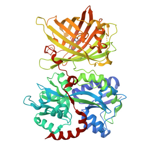
iGABASnFR2 is an improved genetically encoded protein sensor of GABA.
Kolb, I., Hasseman, J.P., Matsumoto, A., Jensen, T.P., Kopach, O., Arthur, B.J., Zhang, Y., Tsang, A., Reep, D., Tsegaye, G., Zheng, J., Patel, R.H., Looger, L.L., Marvin, J.S., Korff, W.L., Rusakov, D.A., Yonehara, K., Turner, G.C.(2026) Elife 14
- PubMed: 41848771 Search on PubMedSearch on PubMed Central
- DOI: https://doi.org/10.7554/eLife.108319
- Primary Citation Related Structures:
9D57 - PubMed Abstract:
Monitoring GABAergic inhibition in the nervous system has been enabled by the development of an intensiometric molecular sensor that directly detects GABA. However, the first generation iGABASnFR exhibits low signal-to-noise and suboptimal kinetics, making in vivo experiments challenging. To improve sensor performance, we targeted several sites in the protein for near-saturation mutagenesis and evaluated the resulting sensor variants in a high-throughput screening system using evoked synaptic release in primary cultured neurons. This identified a sensor variant, iGABASnFR2, with 4.1-fold improved sensitivity and 30% faster rise time, and binding affinity that remained in a range sensitive to changes in GABA concentration at synapses. We also identified sensors with an inverted response, decreasing fluorescence intensity upon GABA binding. We termed the best such negative-going sensor iGABASnFR2n, which can be used to corroborate observations with the positive-going sensor. These improvements yielded a qualitative enhancement of in vivo performance when compared directly to the original sensor. iGABASnFR2 enabled the first measurements of direction-selective GABA release in the retina. In vivo imaging in somatosensory cortex revealed that iGABASnFR2 can report volume-transmitted GABA release following whisker stimulation. Overall, the improved sensitivity and kinetics of iGABASnFR2 make it a more effective tool for imaging GABAergic transmission in intact neural circuits.
- Janelia GENIE Project Team, Janelia Research Campus, Howard Hughes Medical Institute, Ashburn, United States.
Organizational Affiliation: