9D10 | pdb_00009d10

Photoactive Yellow Protein, crystals from PEG, room temperature


Experimental Data Snapshot

  • Method: X-RAY DIFFRACTION
  • Resolution: 1.80 Å
  • R-Value Free: 
    0.198 (Depositor), 0.198 (DCC) 
  • R-Value Work: 
    0.152 (Depositor), 0.152 (DCC) 
  • R-Value Observed: 
    0.155 (Depositor) 

Starting Model: experimental
View more details

wwPDB Validation 3D Report Full Report

Validation slider image for 9D10

Ligand Structure Quality Assessment 


This is version 1.1 of the entry. See complete history

Literature

Exploiting fourth-generation synchrotron radiation for enzyme and photoreceptor characterization.

Malla, T.N.Muniyappan, S.Menendez, D.Ogukwe, F.Dale, A.N.Clayton, J.D.Weatherall, D.D.Karki, P.Dangi, S.Mandella, V.Pacheco, A.A.Stojkovic, E.A.Rose, S.L.Orlans, J.Basu, S.de Sanctis, D.Schmidt, M.

(2025) IUCrJ 12: 36-48

  • DOI: https://doi.org/10.1107/S2052252524010868
  • Primary Citation Related Structures: 
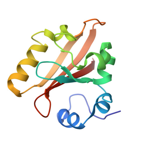
    9CUF, 9D10, 9D2H

  • PubMed Abstract: 

    The upgrade of the European Synchrotron Radiation Facility (ESRF) in Grenoble, France to an Extremely Brilliant Source (EBS) is expected to enable time-resolved synchrotron serial crystallography (SSX) experiments with sub-millisecond time resolution. ID29 is a new beamline dedicated to SSX experiments at ESRF-EBS. Here, we report experiments emerging from the initial phase of user operation at ID29. We first used microcrystals of photoactive yellow protein as a model system to exploit the potential of microsecond pulses for SSX. Subsequently, we investigated microcrystals of cytochrome c nitrite reductase (ccNiR) with microsecond X-ray pulses. CcNiR is a decaheme protein that is ideal for the investigation of radiation damage at the various heme-iron sites. Finally, we performed a proof-of-concept subsecond time-resolved SSX experiment by photoactivating microcrystals of a myxobacterial phytochrome.


  • Organizational Affiliation
    • Department of Physics, University of Wisconsin-Milwaukee, Milwaukee, USA.

Macromolecule Content 

  • Total Structure Weight: 14.05 kDa 
  • Atom Count: 1,066 
  • Modeled Residue Count: 125 
  • Deposited Residue Count: 125 
  • Unique protein chains: 1

Macromolecules

Find similar proteins by:|  3D Structure
Entity ID: 1
MoleculeChains  Sequence LengthOrganismDetailsImage
Photoactive yellow protein125Halorhodospira halophilaMutation(s): 0 
Gene Names: pyp
UniProt
Find proteins for P16113 (Halorhodospira halophila)
Explore P16113 
Go to UniProtKB:  P16113
Entity Groups
Sequence Clusters30% Identity50% Identity70% Identity90% Identity95% Identity100% Identity
UniProt GroupP16113
Sequence Annotations
Expand
Reference Sequence

Small Molecules

Ligands 1 Unique
IDChains Name / Formula / InChI Key2D Diagram3D Interactions
HC4
(Subject of Investigation/LOI)

Query on HC4


Download:Ideal Coordinates CCD File
B [auth A]4'-HYDROXYCINNAMIC ACID
C9 H8 O3
NGSWKAQJJWESNS-ZZXKWVIFSA-N

Experimental Data & Validation

Experimental Data

  • Method: X-RAY DIFFRACTION
  • Resolution: 1.80 Å
  • R-Value Free:  0.198 (Depositor), 0.198 (DCC) 
  • R-Value Work:  0.152 (Depositor), 0.152 (DCC) 
  • R-Value Observed: 0.155 (Depositor) 
Space Group: P 65
Unit Cell:
Length ( Å )Angle ( ˚ )
a = 41.36α = 90
b = 41.36β = 90
c = 118.87γ = 120
Software Package:
Software NamePurpose
PHENIXrefinement
CrystFELdata reduction
CrystFELdata scaling
PHASERphasing

Structure Validation

View Full Validation Report


Ligand Structure Quality Assessment 


Entry History 

& Funding Information

Deposition Data


Funding OrganizationLocationGrant Number
National Science Foundation (NSF, United States)United States1231306
National Science Foundation (NSF, United States)United States2423601

Revision History  (Full details and data files)

  • Version 1.0: 2025-01-15
    Type: Initial release
  • Version 1.1: 2025-01-22
    Changes: Database references