


Structure of the Nudix Hydrolase Rv2985, a Potential Antibiotic Target in M. tuberculosis
Lynch, A.M.To be published.
Experimental Data Snapshot
Starting Model: in silico
View more details
Entity ID: 1 | |||||
|---|---|---|---|---|---|
| Molecule | Chains | Sequence Length | Organism | Details | Image |
| 8-oxo-(d)GTP phosphatase | 341 | Mycobacterium tuberculosis | Mutation(s): 0 Gene Names: mutT1, MT3063 EC: 3.6.1.69 | 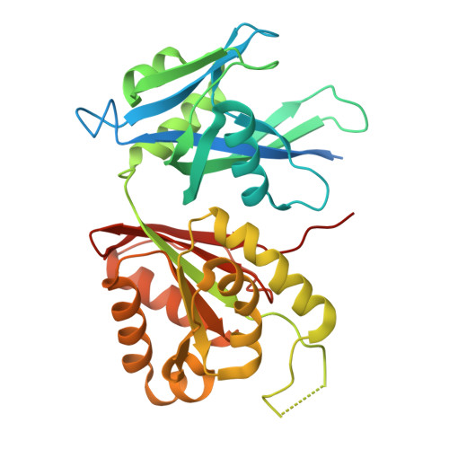 | |
UniProt | |||||
Entity Groups | |||||
| Sequence Clusters | 30% Identity50% Identity70% Identity90% Identity95% Identity100% Identity | ||||
| UniProt Group | P9WIY3 | ||||
Sequence AnnotationsExpand | |||||
Reference Sequence | |||||
| Ligands 2 Unique | |||||
|---|---|---|---|---|---|
| ID | Chains | Name / Formula / InChI Key | 2D Diagram | 3D Interactions | |
| ATP (Subject of Investigation/LOI) Download:Ideal Coordinates CCD File | C [auth A] | ADENOSINE-5'-TRIPHOSPHATE C10 H16 N5 O13 P3 ZKHQWZAMYRWXGA-KQYNXXCUSA-N |  | ||
| SO4 Download:Ideal Coordinates CCD File | D [auth A] E [auth A] F [auth A] G [auth A] H [auth A] | SULFATE ION O4 S QAOWNCQODCNURD-UHFFFAOYSA-L |  | ||
| Length ( Å ) | Angle ( ˚ ) |
|---|---|
| a = 119.005 | α = 90 |
| b = 119.005 | β = 90 |
| c = 115.211 | γ = 90 |
| Software Name | Purpose |
|---|---|
| PHENIX | refinement |
| Coot | model building |
| autoPROC | data scaling |
| PHENIX | phasing |
| Aimless | data scaling |
| pointless | data scaling |
| STARANISO | data scaling |
| XDS | data reduction |
| Funding Organization | Location | Grant Number |
|---|---|---|
| Other private | United States | -- |
| Other private | United States | -- |


