9CV1 | pdb_00009cv1

Crystal structure of the metallo-beta-lactamase VIM-15 with L-captopril


Experimental Data Snapshot

  • Method: X-RAY DIFFRACTION
  • Resolution: 1.29 Å
  • R-Value Free: 
    0.197 (Depositor), 0.197 (DCC) 
  • R-Value Work: 
    0.174 (Depositor), 0.174 (DCC) 
  • R-Value Observed: 
    0.175 (Depositor) 

Starting Model: experimental
View more details

wwPDB Validation 3D Report Full Report

Validation slider image for 9CV1

Ligand Structure Quality Assessment 


This is version 1.0 of the entry. See complete history

Literature

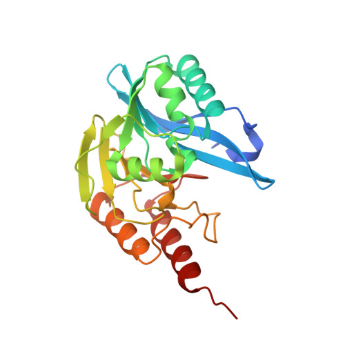
Mutation of an active site-adjacent residue in VIM indirectly dictates interactions with and blunts inhibition by D-captopril.

Silwal, S.B.Wamsley, B.Wang, Z.Gung, B.W.Nix, J.C.Page, R.C.

(2025) J Inorg Biochem 271: 112975-112975

  • DOI: https://doi.org/10.1016/j.jinorgbio.2025.112975
  • Primary Citation Related Structures: 
    9CV1, 9CV2, 9CV3, 9CV4, 9CV5

  • PubMed Abstract: 

    Activity assays and X-ray crystallographic studies were undertaken to elucidate the inhibitory mechanism of captopril stereoisomers on Verona integron-encoded metallo-β-lactamases, specifically VIM-20, VIM-31, and VIM-15. All three VIM-2-like variants (VIM-20, VIM-31, and VIM-15) and VIM-2 expressed in Escherichia coli exhibited catalytic activity with comparable steady-state kinetic parameters. Among the tested thiol drugs (L- and D-captopril, D,L-thiorphan, and 2,3-dimercaprol), IC 50 analyses indicated that D-captopril and 2,3-dimercaprol were more potent inhibitors against the VIM enzymes examined in this study. Notably, the IC 50 value of D-captopril against VIM-31 was an exception, closely resembling that of L-captopril. To elucidate this exceptional inhibitory potency of D-captopril and its binding mode in the active site of VIM-31, high-resolution crystal structures of VIM-20, VIM-31, and VIM-15 in complex with both L- and D-captopril are reported. These findings will help evaluate whether the identified potent inhibitor D-captopril could be further developed as a pan inhibitor targeting the VIM-family enzymes.


  • Organizational Affiliation
    • Department of Chemistry and Biochemistry, Miami University, Oxford, OH, USA.

Macromolecule Content 

  • Total Structure Weight: 26.22 kDa 
  • Atom Count: 1,994 
  • Modeled Residue Count: 234 
  • Deposited Residue Count: 243 
  • Unique protein chains: 1

Macromolecules

Find similar proteins by:|  3D Structure
Entity ID: 1
MoleculeChains  Sequence LengthOrganismDetailsImage
Metallo-beta-lactamase type 2243Pseudomonas aeruginosaMutation(s): 0 
Gene Names: blaVIM-15
EC: 3.5.2.6
UniProt
Find proteins for A0A0F7KYQ8 (Pseudomonas aeruginosa (strain ATCC 15692 / DSM 22644 / CIP 104116 / JCM 14847 / LMG 12228 / 1C / PRS 101 / PAO1))
Explore A0A0F7KYQ8 
Go to UniProtKB:  A0A0F7KYQ8
Entity Groups
Sequence Clusters30% Identity50% Identity70% Identity90% Identity95% Identity100% Identity
UniProt GroupA0A0F7KYQ8
Sequence Annotations
Expand
Reference Sequence

Experimental Data & Validation

Experimental Data

  • Method: X-RAY DIFFRACTION
  • Resolution: 1.29 Å
  • R-Value Free:  0.197 (Depositor), 0.197 (DCC) 
  • R-Value Work:  0.174 (Depositor), 0.174 (DCC) 
  • R-Value Observed: 0.175 (Depositor) 
Space Group: I 2 2 2
Unit Cell:
Length ( Å )Angle ( ˚ )
a = 67.75α = 90
b = 77.51β = 90
c = 79.26γ = 90
Software Package:
Software NamePurpose
PHENIXrefinement
PHENIXrefinement
XDSdata reduction
XDSdata scaling
PHASERphasing

Structure Validation

View Full Validation Report


Ligand Structure Quality Assessment 


Entry History 

& Funding Information

Deposition Data


Funding OrganizationLocationGrant Number
National Institutes of Health/National Institute of General Medical Sciences (NIH/NIGMS)United States128595

Revision History  (Full details and data files)

  • Version 1.0: 2025-07-02
    Type: Initial release