9CU8 | pdb_00009cu8

Crystal Structure of TNFR2 TNFR2_mb1 Complex


Experimental Data Snapshot

  • Method: X-RAY DIFFRACTION
  • Resolution: 1.96 Å
  • R-Value Free: 
    0.223 (Depositor), 0.227 (DCC) 
  • R-Value Work: 
    0.183 (Depositor) 
  • R-Value Observed: 
    0.187 (Depositor) 

Starting Model: in silico
View more details

wwPDB Validation 3D Report Full Report

Validation slider image for 9CU8

This is version 1.1 of the entry. See complete history

Literature

Target-conditioned diffusion generates potent TNFR superfamily antagonists and agonists.

Glogl, M.Krishnakumar, A.Ragotte, R.J.Goreshnik, I.Coventry, B.Bera, A.K.Kang, A.Joyce, E.Ahn, G.Huang, B.Yang, W.Chen, W.Sanchez, M.G.Koepnick, B.Baker, D.

(2024) Science 386: 1154-1161

  • DOI: https://doi.org/10.1126/science.adp1779
  • Primary Citation Related Structures: 
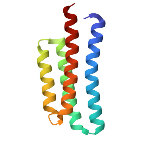
    9CU8

  • PubMed Abstract: 

    Despite progress in designing protein-binding proteins, the shape matching of designs to targets is lower than in many native protein complexes, and design efforts have failed for the tumor necrosis factor receptor 1 (TNFR1) and other protein targets with relatively flat and polar surfaces. We hypothesized that free diffusion from random noise could generate shape-matched binders for challenging targets and tested this approach on TNFR1. We obtain designs with low picomolar affinity whose specificity can be completely switched to other family members using partial diffusion. Designs function as antagonists or as superagonists when presented at higher valency for OX40 and 4-1BB. The ability to design high-affinity and high-specificity antagonists and agonists for pharmacologically important targets in silico presages a coming era in protein design in which binders are made by computation rather than immunization or random screening approaches.


  • Organizational Affiliation
    • Department of Biochemistry, University of Washington, Seattle, WA, USA.

Macromolecule Content 

  • Total Structure Weight: 32.08 kDa 
  • Atom Count: 2,363 
  • Modeled Residue Count: 285 
  • Deposited Residue Count: 296 
  • Unique protein chains: 2

Macromolecules

Find similar proteins by:|  3D Structure
Entity ID: 1
MoleculeChains  Sequence LengthOrganismDetailsImage
TNFR2_mb1106synthetic constructMutation(s): 0 
Find similar proteins by:|  3D Structure
Entity ID: 2
MoleculeChains  Sequence LengthOrganismDetailsImage
Tumor necrosis factor receptor superfamily member 1B190Homo sapiensMutation(s): 0 
Gene Names: TNFRSF1BTNFBRTNFR2
UniProt & NIH Common Fund Data Resources
Find proteins for P20333 (Homo sapiens)
Explore P20333 
Go to UniProtKB:  P20333
PHAROS:  P20333
GTEx:  ENSG00000028137 
Entity Groups
Sequence Clusters30% Identity50% Identity70% Identity90% Identity95% Identity100% Identity
UniProt GroupP20333
Sequence Annotations
Expand
Reference Sequence

Experimental Data & Validation

Experimental Data

  • Method: X-RAY DIFFRACTION
  • Resolution: 1.96 Å
  • R-Value Free:  0.223 (Depositor), 0.227 (DCC) 
  • R-Value Work:  0.183 (Depositor) 
  • R-Value Observed: 0.187 (Depositor) 
Space Group: P 21 21 21
Unit Cell:
Length ( Å )Angle ( ˚ )
a = 35.071α = 90
b = 74.621β = 90
c = 104.973γ = 90
Software Package:
Software NamePurpose
PHENIXrefinement
XDSdata reduction
XSCALEdata scaling
PHASERphasing

Structure Validation

View Full Validation Report


Entry History 

& Funding Information

Deposition Data


Funding OrganizationLocationGrant Number
Howard Hughes Medical Institute (HHMI)United States--

Revision History  (Full details and data files)

  • Version 1.0: 2024-12-04
    Type: Initial release
  • Version 1.1: 2024-12-18
    Changes: Database references