9CQ8 | pdb_00009cq8

Dimeric 9C2 gamma delta TCR extracellular domain bound by Fab 2


Experimental Data Snapshot

  • Method: ELECTRON MICROSCOPY
  • Resolution: 3.45 Å
  • Aggregation State: PARTICLE 
  • Reconstruction Method: SINGLE PARTICLE 

wwPDB Validation 3D Report Full Report

Validation slider image for 9CQ8

This is version 1.0 of the entry. See complete history

Literature

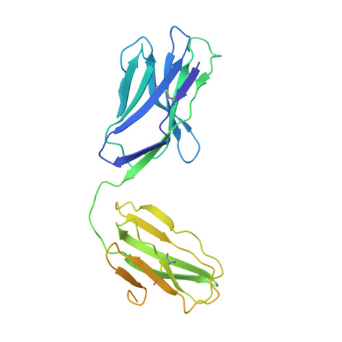
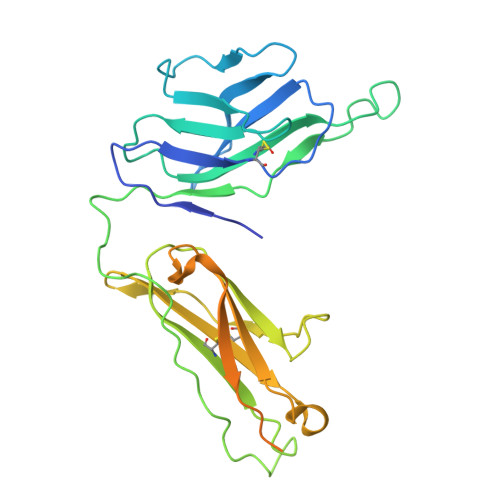
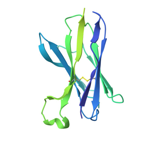
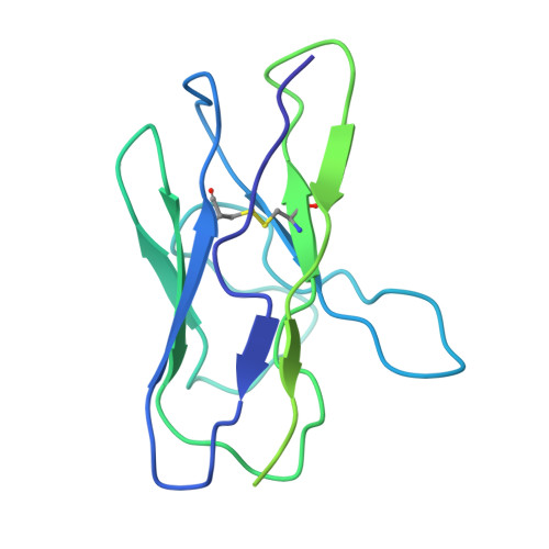
Structural characterization of two gamma delta TCR/CD3 complexes.

Hoque, M.Grigg, J.B.Ramlall, T.Jones, J.McGoldrick, L.L.Lin, J.C.Olson, W.C.Smith, E.Franklin, M.C.Zhang, T.Saotome, K.

(2025) Nat Commun 16: 318-318

  • DOI: https://doi.org/10.1038/s41467-024-55467-5
  • Primary Citation Related Structures: 
    9CQ4, 9CQ7, 9CQ8, 9CQL

  • PubMed Abstract: 

    The T-cell receptor (TCR)/CD3 complex plays an essential role in the immune response and is a key player in cancer immunotherapies. There are two classes of TCR/CD3 complexes, defined by their TCR chain usage (αβ or γδ). Recently reported structures have revealed the organization of the αβ TCR/CD3 complex, but similar studies regarding the γδ TCR/CD3 complex have lagged behind. Here, we report cryoelectron microscopy (cryoEM) structural analysis of two γδ TCRs, G115 (Vγ9 Vδ2) and 9C2 (Vγ5 Vδ1), in complex with CD3 subunits. Our results show that the overall subunit organization of the γδ TCR/CD3 complexes is similar to αβ TCRs. However, both γδ TCRs display highly mobile extracellular domains (ECDs), unlike αβ TCRs, which have TCR ECDs that are rigidly coupled to its transmembrane (TM) domains. We corroborate this finding in cells by demonstrating that a γδ T-cell specific antibody can bind a site that would be inaccessible in the more rigid αβ TCR/CD3 complex. Furthermore, we observed that the Vγ5 Vδ1 complex forms a TCR γ5 chain-mediated dimeric species whereby two TCR/CD3 complexes are assembled. Collectively, these data shed light on γδ TCR/CD3 complex formation and may aid the design of γδ TCR-based therapies.


  • Organizational Affiliation
    • Regeneron Pharmaceuticals, Inc., Tarrytown, NY, 10591, USA. mohammed.hoque@regeneron.com.

Macromolecule Content 

  • Total Structure Weight: 227.24 kDa 
  • Atom Count: 10,480 
  • Modeled Residue Count: 1,306 
  • Deposited Residue Count: 2,024 
  • Unique protein chains: 4

Macromolecules

Find similar proteins by:|  3D Structure
Entity ID: 1
MoleculeChains  Sequence LengthOrganismDetailsImage
9C2 TCR delta chainA,
E [auth D]
280Homo sapiensMutation(s): 0 
Find similar proteins by:|  3D Structure
Entity ID: 2
MoleculeChains  Sequence LengthOrganismDetailsImage
9C2 TCR gamma chain
B, F
294Homo sapiensMutation(s): 0 
Find similar proteins by:|  3D Structure
Entity ID: 3
MoleculeChains  Sequence LengthOrganismDetailsImage
anti TCR variable delta 1 Fab heavy chain
C, G
225Mus musculusMutation(s): 0 
Find similar proteins by:|  3D Structure
Entity ID: 4
MoleculeChains  Sequence LengthOrganismDetailsImage
anti TCR variable delta 1 Fab light chainD [auth E],
H
213Mus musculusMutation(s): 0 

Oligosaccharides

Help  
Entity ID: 5
MoleculeChains Length2D Diagram GlycosylationD Interactions
beta-D-mannopyranose-(1-4)-2-acetamido-2-deoxy-beta-D-glucopyranose-(1-4)-2-acetamido-2-deoxy-beta-D-glucopyranose
I, J, K, L
3N-Glycosylation
Glycosylation Resources
GlyTouCan: G15407YE
GlyCosmos: G15407YE
GlyGen: G15407YE

Experimental Data & Validation

Experimental Data

  • Method: ELECTRON MICROSCOPY
  • Resolution: 3.45 Å
  • Aggregation State: PARTICLE 
  • Reconstruction Method: SINGLE PARTICLE 
EM Software:
TaskSoftware PackageVersion
MODEL REFINEMENTPHENIX1.21.1_5286

Structure Validation

View Full Validation Report


Entry History 

& Funding Information

Deposition Data


Funding OrganizationLocationGrant Number
Other privateUnited States--

Revision History  (Full details and data files)

  • Version 1.0: 2025-01-15
    Type: Initial release