9CCF | pdb_00009ccf

Crystal Structure of DA7-2


Experimental Data Snapshot

  • Method: X-RAY DIFFRACTION
  • Resolution: 4.00 Å
  • R-Value Free: 
    0.296 (Depositor), 0.297 (DCC) 
  • R-Value Work: 
    0.236 (Depositor), 0.236 (DCC) 
  • R-Value Observed: 
    0.242 (Depositor) 

Starting Model: in silico
View more details

wwPDB Validation 3D Report Full Report

Validation slider image for 9CCF

This is version 1.0 of the entry. See complete history

Literature

Design of intrinsically disordered region binding proteins.

Wu, K.Jiang, H.Hicks, D.R.Liu, C.Muratspahic, E.Ramelot, T.A.Liu, Y.McNally, K.Kenny, S.Mihut, A.Gaur, A.Coventry, B.Chen, W.Bera, A.K.Kang, A.Gerben, S.Lamb, M.Y.Murray, A.Li, X.Kennedy, M.A.Yang, W.Song, Z.Schober, G.Brierley, S.M.O'Neill, J.Gelb, M.H.Montelione, G.T.Derivery, E.Baker, D.

(2025) Science 389: eadr8063-eadr8063

  • DOI: https://doi.org/10.1126/science.adr8063
  • Primary Citation Related Structures: 
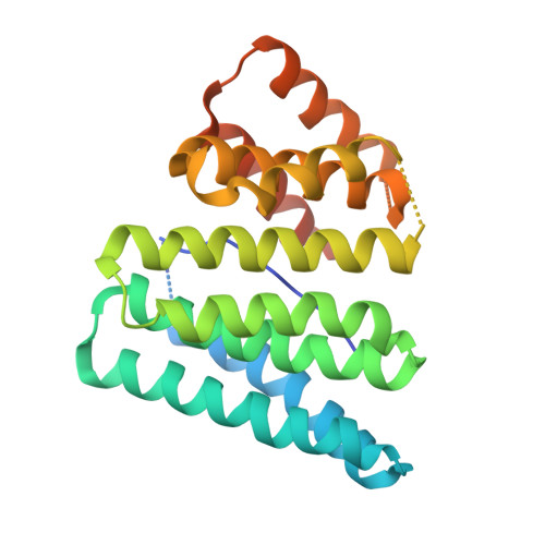
    9CCE, 9CCF

  • PubMed Abstract: 

    Intrinsically disordered proteins and peptides play key roles in biology, but a lack of defined structures and high variability in sequence and conformational preferences have made targeting such systems challenging. We describe a general approach for designing proteins that bind intrinsically disordered protein regions in diverse extended conformations with side chains fitting into complementary binding pockets. We used the approach to design binders for 39 highly diverse unstructured targets, including polar targets, and obtained designs with 100-picomolar to 100-nanomolar affinities in 34 cases, testing ~22 designs per target. The designs function in cells and as detection reagents and are specific for their intended targets in all-by-all binding experiments. Our approach is a major step toward a general solution to the intrinsically disordered protein and peptide recognition problem.


  • Organizational Affiliation
    • Department of Biochemistry, University of Washington, Seattle, WA, USA.

Macromolecule Content 

  • Total Structure Weight: 56.36 kDa 
  • Atom Count: 3,221 
  • Modeled Residue Count: 394 
  • Deposited Residue Count: 482 
  • Unique protein chains: 1

Macromolecules

Find similar proteins by:|  3D Structure
Entity ID: 1
MoleculeChains  Sequence LengthOrganismDetailsImage
DA7_2
A, B
241Homo sapienssynthetic construct
This entity is chimeric
Mutation(s): 0 
Gene Names: PDYN
UniProt & NIH Common Fund Data Resources
Find proteins for P01213 (Homo sapiens)
Explore P01213 
Go to UniProtKB:  P01213
PHAROS:  P01213
GTEx:  ENSG00000101327 
Entity Groups
Sequence Clusters30% Identity50% Identity70% Identity90% Identity95% Identity100% Identity
UniProt GroupP01213
Sequence Annotations
Expand
Reference Sequence

Experimental Data & Validation

Experimental Data

  • Method: X-RAY DIFFRACTION
  • Resolution: 4.00 Å
  • R-Value Free:  0.296 (Depositor), 0.297 (DCC) 
  • R-Value Work:  0.236 (Depositor), 0.236 (DCC) 
  • R-Value Observed: 0.242 (Depositor) 
Space Group: P 1 21 1
Unit Cell:
Length ( Å )Angle ( ˚ )
a = 43.423α = 90
b = 67.214β = 96.746
c = 68.361γ = 90
Software Package:
Software NamePurpose
PHENIXrefinement
XDSdata reduction
XSCALEdata scaling
PHASERphasing

Structure Validation

View Full Validation Report


Entry History 

& Funding Information

Deposition Data


Funding OrganizationLocationGrant Number
Howard Hughes Medical Institute (HHMI)United States--
National Institutes of Health/National Cancer Institute (NIH/NCI)United States--

Revision History  (Full details and data files)

  • Version 1.0: 2025-08-13
    Type: Initial release