9C5A

AP-3 Arf1 dimeric interface, focused refinement


Experimental Data Snapshot

  • Method: ELECTRON MICROSCOPY
  • Resolution: 4.20 Å
  • Aggregation State: PARTICLE 
  • Reconstruction Method: SINGLE PARTICLE 

Starting Model: in silico
View more details

wwPDB Validation   3D Report Full Report


This is version 1.1 of the entry. See complete history


Literature

A structure-based mechanism for initiation of AP-3 coated vesicle formation.

Begley, M.Aragon, M.Baker, R.W.

(2024) Proc Natl Acad Sci U S A 121: e2411974121-e2411974121

  • DOI: https://doi.org/10.1073/pnas.2411974121
  • Primary Citation of Related Structures:  
    9C58, 9C59, 9C5A, 9C5B, 9C5C

  • PubMed Abstract: 

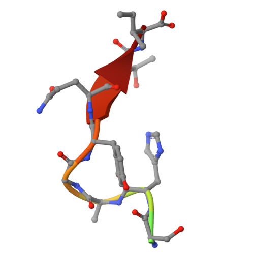
    Adaptor protein complex-3 (AP-3) mediates cargo sorting from endosomes to lysosomes and lysosome-related organelles. Recently, it was shown that AP-3 adopts a constitutively open conformation compared to the related AP-1 and AP-2 coat complexes, which are inactive until undergoing large conformational changes upon membrane recruitment. How AP-3 is regulated is therefore an open question. To understand the mechanism of AP-3 membrane recruitment and activation, we reconstituted human AP-3 and determined multiple structures in the soluble and membrane-bound states using electron cryo-microscopy. Similar to yeast AP-3, human AP-3 is in a constitutively open conformation. To reconstitute AP-3 activation by adenosine di-phosphate (ADP)-ribosylation factor 1 (Arf1), a small guanosine tri-phosphate (GTP)ase, we used lipid nanodiscs to build Arf1-AP-3 complexes on membranes and determined three structures showing the stepwise conformational changes required for formation of AP-3 coated vesicles. First, membrane recruitment is driven by one of two predicted Arf1 binding sites, which flexibly tethers AP-3 to the membrane. Second, cargo binding causes AP-3 to adopt a fixed position and rigidifies the complex, which stabilizes binding for a second Arf1 molecule. Finally, binding of the second Arf1 molecule provides the template for AP-3 dimerization, providing a glimpse into the first step of coat polymerization. We propose coat polymerization only occurs after cargo engagement, thereby linking cargo sorting with assembly of higher-order coat structures. Additionally, we provide evidence for two amphipathic helices in AP-3, suggesting that AP-3 contributes to membrane deformation during coat assembly. In total, these data provide evidence for the first stages of AP-3-mediated vesicle coat assembly.


  • Organizational Affiliation

    Department of Biochemistry and Biophysics, University of North Carolina at Chapel Hill School of Medicine, Chapel Hill, NC 27599.


Macromolecules
Find similar proteins by:  (by identity cutoff)  |  3D Structure
Entity ID: 1
MoleculeChains Sequence LengthOrganismDetailsImage
AP-3 complex subunit beta-1A [auth B],
E [auth b]
684Homo sapiensMutation(s): 0 
Gene Names: AP3B1ADTB3A
UniProt & NIH Common Fund Data Resources
Find proteins for O00203 (Homo sapiens)
Explore O00203 
Go to UniProtKB:  O00203
PHAROS:  O00203
GTEx:  ENSG00000132842 
Entity Groups  
Sequence Clusters30% Identity50% Identity70% Identity90% Identity95% Identity100% Identity
UniProt GroupO00203
Sequence Annotations
Expand
  • Reference Sequence
Find similar proteins by:  (by identity cutoff)  |  3D Structure
Entity ID: 2
MoleculeChains Sequence LengthOrganismDetailsImage
AP-3 complex subunit mu-1B [auth M],
F [auth m]
418Homo sapiensMutation(s): 0 
Gene Names: AP3M1
UniProt & NIH Common Fund Data Resources
Find proteins for Q9Y2T2 (Homo sapiens)
Explore Q9Y2T2 
Go to UniProtKB:  Q9Y2T2
PHAROS:  Q9Y2T2
GTEx:  ENSG00000185009 
Entity Groups  
Sequence Clusters30% Identity50% Identity70% Identity90% Identity95% Identity100% Identity
UniProt GroupQ9Y2T2
Sequence Annotations
Expand
  • Reference Sequence
Find similar proteins by:  (by identity cutoff)  |  3D Structure
Entity ID: 3
MoleculeChains Sequence LengthOrganismDetailsImage
ADP-ribosylation factor 1C,
G [auth c]
182Homo sapiensMutation(s): 1 
Gene Names: ARF1
EC: 3.6.5.2
UniProt & NIH Common Fund Data Resources
Find proteins for P84077 (Homo sapiens)
Explore P84077 
Go to UniProtKB:  P84077
PHAROS:  P84077
GTEx:  ENSG00000143761 
Entity Groups  
Sequence Clusters30% Identity50% Identity70% Identity90% Identity95% Identity100% Identity
UniProt GroupP84077
Sequence Annotations
Expand
  • Reference Sequence

Find similar proteins by:  Sequence   |   3D Structure  

Entity ID: 4
MoleculeChains Sequence LengthOrganismDetailsImage
Lysosome-associated membrane glycoprotein 1D [auth Y],
H [auth y]
12Homo sapiensMutation(s): 0 
UniProt & NIH Common Fund Data Resources
Find proteins for P11279 (Homo sapiens)
Explore P11279 
Go to UniProtKB:  P11279
PHAROS:  P11279
GTEx:  ENSG00000185896 
Entity Groups  
Sequence Clusters30% Identity50% Identity70% Identity90% Identity95% Identity100% Identity
UniProt GroupP11279
Sequence Annotations
Expand
  • Reference Sequence
Experimental Data & Validation

Experimental Data

  • Method: ELECTRON MICROSCOPY
  • Resolution: 4.20 Å
  • Aggregation State: PARTICLE 
  • Reconstruction Method: SINGLE PARTICLE 
EM Software:
TaskSoftware PackageVersion
RECONSTRUCTIONcryoSPARC4.4.1
MODEL REFINEMENTRosetta3.12
MODEL REFINEMENTPHENIX1.21.1

Structure Validation

View Full Validation Report


Entry History & Funding Information

Deposition Data


Funding OrganizationLocationGrant Number
National Institutes of Health/National Institute of General Medical Sciences (NIH/NIGMS)United StatesR35 GM150960

Revision History  (Full details and data files)

  • Version 1.0: 2024-12-18
    Type: Initial release
  • Version 1.1: 2025-03-05
    Changes: Data collection, Database references