9C42 | pdb_00009c42

Structure of human MR1-ellagic acid in complex with human MAIT A-F7 TCR


Experimental Data Snapshot

  • Method: X-RAY DIFFRACTION
  • Resolution: 2.69 Å
  • R-Value Free: 
    0.246 (Depositor), 0.246 (DCC) 
  • R-Value Work: 
    0.194 (Depositor), 0.197 (DCC) 
  • R-Value Observed: 
    0.196 (Depositor) 

Starting Model: experimental
View more details

wwPDB Validation 3D Report Full Report

Validation slider image for 9C42

Ligand Structure Quality Assessment 


This is version 1.2 of the entry. See complete history

Literature

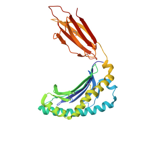
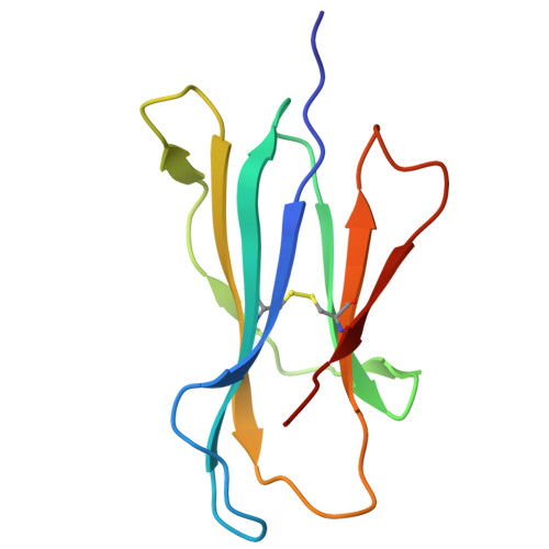
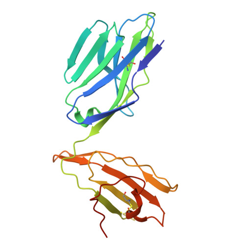
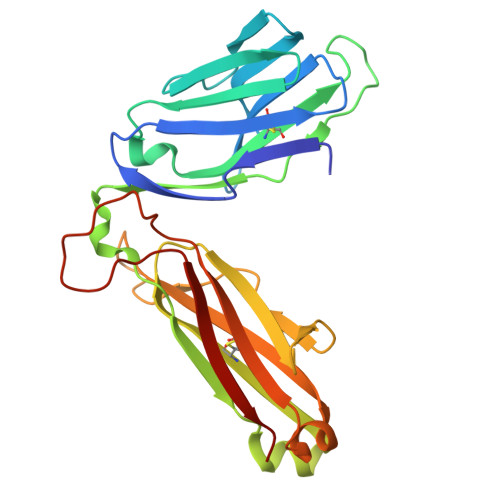
Ellagic acid metabolism as a source of dietary MR1 ligands.

O'Sullivan, D.Wang, C.J.H.Tang, J.S.Stephens, R.Faulkner, S.Gell, K.Li, Y.Cait, A.McInerney, M.Purcell, A.W.Rossjohn, J.Le Nours, J.Gasser, O.

(2026) J Immunol 215

  • DOI: https://doi.org/10.1093/jimmun/vkaf346
  • Primary Citation Related Structures: 
    9C42

  • PubMed Abstract: 

    MR1 is an major histocompatibility complex class I-like molecule that presents small molecule metabolites to MR1-restricted T cells that include a major population of highly conserved T cells known as mucosal-associated invariant T (MAIT) cells. MAIT cells recognize bacterial riboflavin pathway-derived neoantigens and are being attributed an increasing number of immune and homeostatic functions. However, the chemical breadth and diversity of MR1-restricted ligands remain to be fully elucidated. Due to the largely (poly)cyclic structure of known MR1 ligands, we aimed to identify MR1 ligands from a library of dietary phenolic metabolites. Competitive MAIT cell inhibition assays using both cell lines and primary cells isolated from human blood identified gut microbial metabolites of ellagitannins that include ellagic acid (EA), urolithin D (UroD), and UroM5 as potential MR1 ligands. Fluorescence polarization binding assays demonstrated that EA, UroM5, UroC, and UroB bound to MR1, and we provide a structural basis for EA presentation by MR1. Overall, our findings indicate that EA metabolism provides dietary MR1 ligands that inhibit T cell receptor-dependent MAIT cell activation.


  • Organizational Affiliation
    • Malaghan Institute of Medical Research, Wellington, New Zealand.

Macromolecule Content 

  • Total Structure Weight: 189.27 kDa 
  • Atom Count: 12,686 
  • Modeled Residue Count: 1,611 
  • Deposited Residue Count: 1,642 
  • Unique protein chains: 4

Macromolecules

Find similar proteins by:|  3D Structure
Entity ID: 1
MoleculeChains  Sequence LengthOrganismDetailsImage
Major histocompatibility complex class I-related gene protein
A, C
271Homo sapiensMutation(s): 0 
Gene Names: MR1
UniProt & NIH Common Fund Data Resources
Find proteins for Q95460 (Homo sapiens)
Explore Q95460 
Go to UniProtKB:  Q95460
PHAROS:  Q95460
GTEx:  ENSG00000153029 
Entity Groups
Sequence Clusters30% Identity50% Identity70% Identity90% Identity95% Identity100% Identity
UniProt GroupQ95460
Sequence Annotations
Expand
Reference Sequence
Find similar proteins by:|  3D Structure
Entity ID: 2
MoleculeChains  Sequence LengthOrganismDetailsImage
Beta-2-microglobulin
B, F
100Homo sapiensMutation(s): 0 
Gene Names: B2MCDABP0092HDCMA22P
UniProt & NIH Common Fund Data Resources
Find proteins for P61769 (Homo sapiens)
Explore P61769 
Go to UniProtKB:  P61769
PHAROS:  P61769
GTEx:  ENSG00000166710 
Entity Groups
Sequence Clusters30% Identity50% Identity70% Identity90% Identity95% Identity100% Identity
UniProt GroupP61769
Sequence Annotations
Expand
Reference Sequence
Find similar proteins by:|  3D Structure
Entity ID: 3
MoleculeChains  Sequence LengthOrganismDetailsImage
TRA@ protein
D, G
204Homo sapiensMutation(s): 0 
Gene Names: TRA@
UniProt
Find proteins for Q6P4G7 (Homo sapiens)
Explore Q6P4G7 
Go to UniProtKB:  Q6P4G7
Entity Groups
Sequence Clusters30% Identity50% Identity70% Identity90% Identity95% Identity100% Identity
UniProt GroupQ6P4G7
Sequence Annotations
Expand
Reference Sequence
Find similar proteins by:|  3D Structure
Entity ID: 4
MoleculeChains  Sequence LengthOrganismDetailsImage
MAIT T-cell receptor beta chain
E, H
246Homo sapiensMutation(s): 0 

Small Molecules

Ligands 5 Unique
IDChains Name / Formula / InChI Key2D Diagram3D Interactions
REF
(Subject of Investigation/LOI)

Query on REF


Download:Ideal Coordinates CCD File
I [auth A],
M [auth C]
2,3,7,8-tetrahydroxychromeno[5,4,3-cde]chromene-5,10-dione
C14 H6 O8
AFSDNFLWKVMVRB-UHFFFAOYSA-N
GOL

Query on GOL


Download:Ideal Coordinates CCD File
Q [auth E],
R [auth F],
S [auth F]
GLYCEROL
C3 H8 O3
PEDCQBHIVMGVHV-UHFFFAOYSA-N
ACY

Query on ACY


Download:Ideal Coordinates CCD File
L [auth A],
O [auth C],
P [auth D]
ACETIC ACID
C2 H4 O2
QTBSBXVTEAMEQO-UHFFFAOYSA-N
CL

Query on CL


Download:Ideal Coordinates CCD File
N [auth C]CHLORIDE ION
Cl
VEXZGXHMUGYJMC-UHFFFAOYSA-M
NA

Query on NA


Download:Ideal Coordinates CCD File
J [auth A],
K [auth A],
T [auth H]
SODIUM ION
Na
FKNQFGJONOIPTF-UHFFFAOYSA-N

Experimental Data & Validation

Experimental Data

  • Method: X-RAY DIFFRACTION
  • Resolution: 2.69 Å
  • R-Value Free:  0.246 (Depositor), 0.246 (DCC) 
  • R-Value Work:  0.194 (Depositor), 0.197 (DCC) 
  • R-Value Observed: 0.196 (Depositor) 
Space Group: C 1 2 1
Unit Cell:
Length ( Å )Angle ( ˚ )
a = 215.81α = 90
b = 70.352β = 104.09
c = 143.434γ = 90
Software Package:
Software NamePurpose
PHENIXrefinement
XDSdata reduction
Aimlessdata scaling
PHENIXphasing

Structure Validation

View Full Validation Report


Ligand Structure Quality Assessment 


Entry History 

& Funding Information

Deposition Data


Funding OrganizationLocationGrant Number
National Health and Medical Research Council (NHMRC, Australia)Australia2008981

Revision History  (Full details and data files)

  • Version 1.0: 2025-06-11
    Type: Initial release
  • Version 1.1: 2025-06-18
    Changes: Derived calculations
  • Version 1.2: 2026-03-11
    Changes: Database references